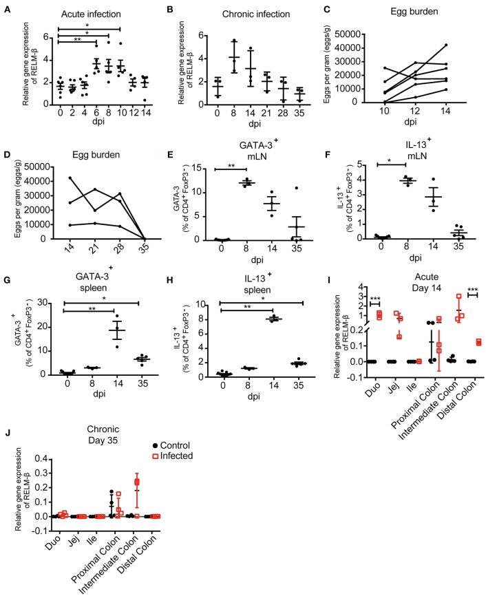
Figure 2.
Immunological and parasitological analysis of murine stool after infection with Heligmosomoides polygyrus. BALB/c mice were infected orally with 200 infectious L3 stage larvae of H. polygyrus. Parameters were measured at different time points and in different regions of the intestine. (A) Relative gene expression of RELM-β in exfoliated intestinal cells during acute infection (day 0−14 dpi). (B) RELM-β gene expression in exfoliated cells until chronicity of infection (day 0−35 dpi), shown as mean ± SD. (C) Fecal egg counts during acute H. polygyrus infection (day 14 dpi). (D) Fecal egg counts during chronic H. polygyrus infection (day 14−35 dpi). Frequency of CD4+ T cells expressing GATA-3 (E) and IL-13 (F) in mesenteric lymph nodes (mLN). (G) Frequencies of CD4+ T cell expressing GATA-3 and IL13 (H) in spleen. (I,J) RELM-β expression in intestinal tissue (duodenum, jejunum, ileum, proximal colon, intermediate colon, and distal colon) at 14 and 35 dpi, respectively. All relative gene expression analysis is compared to β-actin using 10 ng cDNA. Data from (A) is pooled from two independent experiments with n = 4–10 fecal pellets from 4–6 animals. (B) is representative of two independent experiments, n = 3. (C) is pooled from two independent experiments, n = 6. (D–H) is representative of two independent experiments, n = 3. (I,J) is representative of two independent experiments, n = 3–4. (A) shown as mean ± SEM, *P ≤ 0.05, **P ≤ 0.01, and ***P ≤ 0.001. (B–H) shown as mean ± SD; (A–H) Statistical analysis was performed using the Kruskal-Wallis with Dunn's multiple comparison test, (I,J) multiple t tests and corrected for multiple comparisons using the Holm-Sidak method, *P ≤ 0.05, **P ≤ 0.01, and ***P ≤ 0.001. dpi: days post infection.

