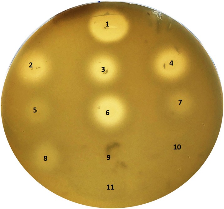
FIGURE 4.

Hyaluronidase activity of S. saccharolyticus. Detection of hyaluronate lyase activity with a hyaluronic acid plate assay. Colonies of S. saccharolyticus strains were point-inoculated onto hyaluronic acid-containing plates and incubated for 48 h under anaerobic conditions. Plates were flushed with 2N acetic acid for 15 min for the detection of hyaluronic acid degradation. Numbers (in brackets the respective S. saccharolyticus subclade): 1, positive control (hyaluronidase from Streptococcus pyogenes); 2, 05B0362 (1); 3, 12B0021 (1); 4, 13T0028 (1); 5, DVP2-17-2406 (2); 6, DVP3-16-6167 (1); 7, DVP4-17-2404 (2); 8, DVP5-16-4677 (2); 9, S. epidermidis FSI; 10, S. epidermidis 14.1.R1; 11, BHCY medium (negative control). The pictures are representative of three independent experiments.
