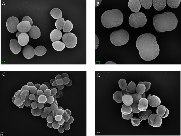
FIGURE 5.
Scanning electron microscopy of S. saccharolyticus. The upper (A,B) and lower (C,D) panels show representative images of S. saccharolyticus 13T0028 (subclade 1) and S. saccharolyticus DVP4-17-2404 (subclade 2), respectively. Images indicate morphological differences between these two strains.

