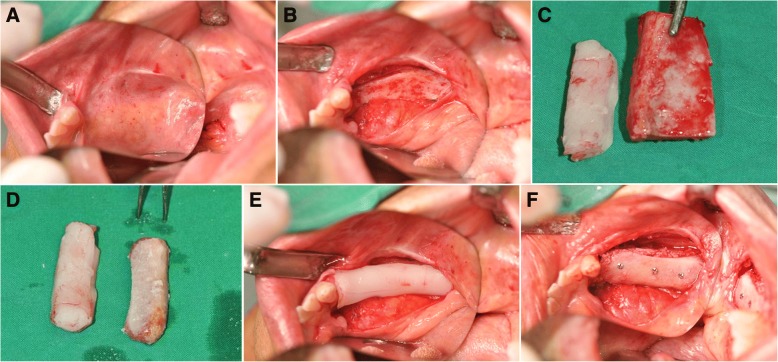
Fig. 2.
Clinical views of patient no. 07 during the bone graft stage showing pre-operative view of the right edentulous posterior region of the mandible (a), elevation of a full thickness mucoperiosteal flap to expose the deficient edentulous area (b), the iliac bone block was modeled to obtain precise adaptation to the recipient region (c, d), and iliac bone block was fixed to the basal bone using titanium microscrews (e, f). All gaps between bone blocks were filled with autogenous particulate bone

