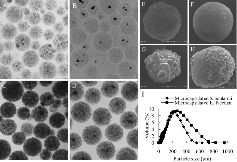
Fig. 1.

The morphology of micro-beads loaded with bacteria and their growth profile. a, b Micro images of freshly prepared micro-beads loaded with S. boulardii or E. faecium. c Micro-beads loaded with S. boulardii after 18 h of incubation in YPD broth with 180-rpm shaking at 30 °C. d Micro-beads loaded with E. faecium after 14 h of incubation in MRS broth at 37 °C. e, f Scanning electron micrograph of micro-beads freshly loaded with S. boulardii or E. faecium. g, h Scanning electron micrograph of micro-beads loaded with S. boulardii or E. faecium, after incubation. i The size distribution of micro-beads loaded with S. boulardii or E. faecium was measured by a laser particle analyzer (n = 3). All micro images show 40 × magnification
