Abstract
Bioassay guided isolation from the leaves of Rauvolfia tetraphylla L. resulted in the isolation and characterization of three compounds of alkaloid in nature namely, Curan-17-oic acid (F1); 18, 19-Secoyohimban (F2) and Reserpiline (F3). Macrofilaricidal activity of three compounds was tested against bovine filarial parasite Setaria cervi using in vitro assays and supported by in silico docking analysis on glutathione-S-transferase (GST) enzyme of Wuchereria bancrofti. All the molecules inhibited GST enzyme to some extent 35.78%, 78.22% and 64.21% respectively. Results were supported by molecular docking studies, which showed docking scores for compound F1 (− 5.14), compound F2 (− 7.19) and compound F3 (− 7.2) on GST enzyme. Thus, in conclusion the in vitro and in silico studies indicated that isolated compounds are promising, inexpensive and widely available natural leads, which can be designed and developed into the macrofilaricidal drugs.
Keywords: Rauvolfia tetraphylla, Setaria cervi, MTT, Glutathione-S-transferase, Macrofilaricidal activity, In-silico docking
Introduction
Filariasis is one of the most important tropical diseases affecting more than 80 countries with 120 million infected and more than 1.3 million people at risk globally. Two-third of the endemic population resides in South-East Asia and one-third lives in India. This is also called a poor man’s disease prevailing in most of the slum areas (Dreyer et al. 1997). Lymphatic filariasis or elephantiasis is the fourth most common cause of disability worldwide. The filarial nematode W. bancrofti accounts for 91% of Lymphatic Filariasis infections, while B. malayi and B. timori are responsible for the remaining 9% in the Southeast Asia region (WHO 2009). The control of human filariasis caused by Brugia malayi, Wuchereria bancrofti and Onchocerca volvulus, currently relies on community-wide mass distribution of ivermectin and albendazole, either individually or in combination with diethylcarbamazine. Unfortunately, these drugs are mainly microfilaricidal rather than macrofilaricidal, which means that repeated treatment is required over many years, and the possibility that resistance to them may develop is a cause for concern (Ardelli et al. 2005; Schwab et al. 2005) but the same when given for a longer duration are full of side effects like nausea, vomiting and head ache.
The antifilarial potential of crude extracts of Rauvolfia tetraphylla plant has been analysed profoundly in our previous studies that signified this plant being a promising source of antifilarial principles (Behera and Bhatnagar 2017). In the present study, R. tetraphylla L. one of the most important medicinal plant species of Apocynaceae family was explored for the evaluation of macrofilaricidal potential and the same was also employed for the isolation of antifilarial principles by targeting bovine filarial parasite Setaria cervi. The in vitro activity of isolated compounds against adult worms of S. cervi, in silico docking on glutathione-S-transferase (GST) enzyme of W. bancrofti parasite model have been discussed here in detail.
Materials and methods
General experimental procedures
Column chromatography was performed by using 60–120 mesh silica gel (Sisco Research Laboratory, India) through a glass column of size 400 × 30 mm. Preparative TLC was carried out on prep. TLC glass plates coated with silica gel GF254 (Merck, Germany). UV spectral data were obtained on a Shimadzu UV-1800 double beam spectrophotometer (Tokyo, Japan). HPLC analysis was done on a Shimadzu—UFLC (Tokyo, Japan) system that includes a C18 column (150 mm × 4.6 mm, 5 µm) with a biocompatible binary pump (LC-20AD) plus a wavelength detector (SPD-M20A). IR spectra of samples in KBr discs were recorded on a Perkin Elmer-Spectrum-GX spectrometer (PerkinElmer, USA) with KBr pellets. GC–MS analysis of isolated compounds was carried out using GCMS-QP 2010 Ultra instrument (Shimadzu, Japan) that includes a headspace auto sampler (AOC-20s) and auto injector (AOC-20i). NMR experiments were carried out on a Bruker Avance III 500 MHz spectrometer (Karlsruhe, Germany) with pulsed field gradient and signals were referenced to the residual solvent signals (CD3OD, at δH 3.31 and δC 49.0 ppm). Melting point detection was done by a digital melting point apparatus (BPL-53A, BP lab solutions, India). TLC was carried out on silica gel 60 F254 TLC sheets (Merck, Germany) and spots were detected by spraying agents like DPPH (0.2% in CH3OH), Vanillin- H2SO4 in EtOH and Dragondroff’s reagent.
Collection of parasites
Setaria cervi, resembles the human bancroftian parasite in its nocturnal periodicity, antigenic pattern and same chemotherapeutic antifilarial drug response, has been used as a model parasite for drug discovery research in filariasis (Kaushal et al. 1987). The adult parasites (S. cervi) were collected from peritoneal cavity of freshly slaughtered buffalo obtained from slaughter house of Nandan Kanan Zoological Park, Bhubaneswar. These adult parasites were washed in PBS (1 ×) and transferred immediately to RPMI-1640 medium supplemented with 5% (v/v) heat-inactivated Fetal bovine serum (Mathew et al. 2002). The adult female parasites measuring average length 80–100 mm and width 0.45 mm were used for the experimental purposes.
Plant sample collection and preparation
The fresh leaves of R. tetraphylla were collected from medicinal germplasm garden, RPRC botanical garden and forest area of Regional Plant Resource Centre, Bhubaneswar, Odisha, India and voucher specimen (RPRC-15010) was deposited to herbarium of Regional Plant Resource Centre, Bhubaneswar, Odisha. The leaves were washed thoroughly in running tap water and shade dried at room temperature. The dried leaves were grinded to make powder; same was used for preparing the solvent extracts.
Extraction and isolation
The crude extracts such as hexane, chloroform, acetone and methanol were prepared by soxhlet extraction technique. The crude methanolic extract (50 g dry weight) was fractionated by liquid–liquid separation technique using solvents such as Hexane, Chloroform and Ethyl acetate according to increasing polarity. The crude plant extract was separately suspended in distilled water using separating funnel (100 ml) and successively partitioned with hexane, chloroform and ethyl acetate in the order of polarity. This afforded four partition fractions of active plant extract. The chloroform fraction (20 g) was subjected to column chromatography over silica gel (60–120 mesh), twenty sub-fractions were collected using mobile phase starting from hexane (100%), dichloromethane, chloroform, ethyl acetate and methanol in the ratio of 100, 90: 10, 70: 30, 50: 50 and 20: 80. Further, the sub-fractions were combined according to their similarity in Rf values determined by TLC in mobile phase toluene: ethyl acetate: methanol (3.6.1) and therefore resulting in five combined sub-fractions. Further purification of sub-fractions was achieved by preparative TLC method (prep. TLC silica gel plates, Merck) using mobile phase toluene: ethyl acetate: methanol (3.6.1) and benzene: ethanol: ammonium hydroxide (90: 10: 1). The compounds 1–3 were examined using standard protocols for the identification of their chemical nature and therefore phytochemical study was carried out by the methods described by Sofowora (1993) and Trease and Evans (1989). Then the compounds 1–3 (40 mg) were filtered by syringe filters (PTFE membrane, pore size 0.45 µm, Axiva) and purity was checked by TLC (Silica gel 60 F254, Merck) and HPLC (Shimadzu) using a C18 column (150 mm × 4.6 mm, 5 µm) with mobile phase CH3OH: C2H3N (60: 40 v/v) (Masoko and Eloff 2007; Malinowska et al. 2005; Panwar and Guru 2011; Kardono et al. 1990).
Compound 1 [Curan-17-oic acid, 19, 20-dihydroxy, methyl ester (19S)]: Amorphous solid; λmax 234, 278.5 and 303.5 nm; melting point: 255–265 °C; IR (KBr) νmax: 3360, 2930, 1687, 1623, 1437, 800, 620 cm−1. GC–MS m/z [M + H] + 358; 1H NMR (500 MHz, CD3OD): δ 1.19 (3H, d, J = 6.2 Hz), 1.63–1.87 (3H, 1.82 (ddd, J = 13.8, 3.0, 2.3 Hz), 1.72 (ddd, J = 13.7, 6.8, 1.7 Hz), 1.71 (ddd, J = 13.7, 7.8, 6.9 Hz)), 1.92 (1H, ddd, J = 13.8, 3.3, 2.5 Hz), 2.39 (1H, dt, J = 3.3, 2.3 Hz), 2.56 (1H, ddd, J = 12.9, 7.8, 6.8 Hz), 2.70 (1H, d, J = 7.9 Hz), 2.88–3.07 (3H, 2.94 (ddd, J = 12.9, 6.9, 1.7 Hz), 3.01 (dd, J = 6.4, 2.3 Hz), 3.04 (d, J = 7.9 Hz)), 3.31 (1H, dd, J = 3.0, 2.5 Hz), 3.65 (3H, s), 3.70–3.80 (2H, 3.74 (d, J = 6.4 Hz), 3.75 (q, J = 6.2 Hz)), 6.54 (1H, td, J = 7.6, 1.1 Hz), 6.69 (1H, ddd, J = 7.8, 1.1, 0.4 Hz), 6.90–6.99 (2H, 6.94 (ddd, J = 7.8, 7.6, 1.5 Hz), 6.93 (ddd, J = 7.6, 1.5, 0.4 Hz)). 13C-NMR (125 MHz, CD3OD): 53.3 (C-7), 62.15 (C-8), 58.06 (C-9), 44.64 (C-10), 34.45 (C-11), 40.95 (C-12), 79.36 (C-13), 39.9 (C-14), 61.71 (C-15), 52.27 (C-16), 137.7 (C-17), 149.89 (C-18), 71.67 (C-19), 172.03 (C-20), 122.5 (C-21), 109.8 (C-22), 17.19 (C-23), 120.92 (C-24), 129.58 (C-25), 52.08 (C-26).
Compound 2 [18,19-Secoyohimban-19-oic acid, 16,17,20,21-tetradehydro-16-(hydroxymethyl)-, methyl ester]: Amorphous solid; λmax 227.5, 303.5 and 401.5 nm; melting point: 240–250 °C; IR (KBr) νmax: 3390, 2927, 1707, 1624, 1210, 750, 470 cm−1.GC–MS m/z [M + H] + 354; 1H NMR (500 MHz, CD3OD): δ 1.94 (3H, d, J = 7.1 Hz), 2.16–2.41 (2H, 2.24 (ddd, J = 13.8, 9.8, 9.3 Hz), 2.35 (ddd, J = 13.8, 4.9, 4.2 Hz)), 3.11–3.26 (2H, 3.18 (ddd, J = 14.1, 8.9, 5.3 Hz), 3.17 (ddd, J = 14.1, 5.4, 1.4 Hz)), 3.42 (1H, dd, J = 9.3, 4.9 Hz), 3.54 (1H, ddd, J = 13.8, 8.9, 5.4 Hz), 3.62–3.73 (4H, 3.70 (s), 3.68 (ddd, J = 13.8, 5.3, 1.4 Hz)), 5.16 (1H, dd, J = 9.8, 4.2 Hz), 6.75 (1H, q, J = 7.1 Hz), 6.92 (1H, ddd, J = 7.8, 7.6, 1.2 Hz), 7.05–7.15 (2H, 7.08 (ddd, J = 7.8, 1.2, 0.5 Hz), 7.10 (ddd, J = 7.8, 7.6, 1.3 Hz)), 7.40 (1H, ddd, J = 7.8, 1.3, 0.5 Hz), 8.44 (1H, s), 9.67 (1H, s). 13C-NMR (125 MHz, CD3OD): δ 58.23 (C-6), 20.50 (C-7), 134.44 (C-8), 38.00 (C-9), 50.80 (C-10), 107.64 (C-11), 21.90 (C-12), 101.80 (C-13), 139.84 (C-14), 127.37 (C-15), 136.07 (C-16), 146.43 (C-17), 117.95 (C-18), 166.60 (C-19), 63.35 (C-20), 110.93 (C-21), 129.64 (C-22), 119.52 (C-23), 121.26 (C-24), 13.88 (C-25), 50.40 (C-26).
Compound 3 [Reserpiline/Oxayohimban-16-carboxylic acid, 16, 17-didehydro-10,11-dimethoxy-19-methyl-, methyl ester, (3-beta, 19-alpha, 20-alpha)-]: Amorphous solid; λmax 228 and 279.5 nm; melting point: 250–260 °C; IR (KBr) νmax: 3407, 2927, 1629, 1620, 1089, 800, 480 cm−1.GC–MS m/z [M + H] + 412 1H NMR (500 MHz, CD3OD): δ 1.18 (3H, d, J = 6.4 Hz), 1.82 (1H, ddd, J = 13.6, 2.6, 2.1 Hz), 1.96 (1H, ddd, J = 13.6, 10.3, 3.7 Hz), 2.17 (1H, dddd, J = 10.3, 4.1, 3.0, 2.8 Hz), 2.46 (1H, dd, J = 10.3, 6.6 Hz), 2.72–2.92 (5H, 2.87 (dd, J = 6.6, 3.0 Hz), 2.83 (ddd, J = 4.1, 3.7, 2.1 Hz), 2.79 (ddd, J = 14.4, 10.0, 3.9 Hz), 2.87 (ddd, J = 14.4, 4.0, 1.9 Hz), 2.84 (ddd, J = 13.8, 10.0, 4.0 Hz)), 3.01 (1H, ddd, J = 13.8, 3.9, 1.9 Hz), 3.54 (3H, s), 3.60 (3H, s), 3.72 (3H, s), 3.91 (1H, dd, J = 10.3, 2.6 Hz), 5.02 (1H, qd, J = 6.4, 2.8 Hz), 6.32 (1H, d, J = 0.5 Hz), 6.38 (1H, d, J = 0.5 Hz), 8.03 (1H, s). 13C NMR (125 MHz, CD3OD): δ 40.73 (C-8), 30.50 (C-9), 60.00 (C-10), 32.63 (C-11), 56.67 (C-12), 73.63 (C-13), 134.37 (C-14), 53.10 (C-15), 107.63 (C-16), 21.23 (C-17), 107.43 (C-18), 128.30 (C-19), 15.84 (C-20), 154.63 (C-21), 136.83 (C-22), 167.43 (C-23), 98.70 (C-24), 93.90 (C-25), 149.61 (C-26), 148.97 (C-27), 51.58 (C-28), 56.13 (C-29), 56.16 (C-30).
Macrofilaricidal assays on model parasite S. cervi
The isolated compounds alone and in combination with Diethylcarbamazine (DEC) were prepared at concentrations 1.0, 0.5 and 0.25 mg/ml in dimethyl sulfoxide (DMSO) to determine the efficacy of the pure compounds on the target organism. In vitro worm motility inhibition and MTT reduction assay was carried out by the standard procedures described earlier (Murthy 1999; Comley et al. 1989; Sahare and Singh 2013; Lakshmi et al. 2010). The experiment was conducted in two replicates with one adult female worm per well in RPMI 1640 media supplemented with 0.5% Fetal Bovine Serum. A set of control worms (DMSO treated) were also maintained along with the treated worms for each doses to calculate inhibition percentage. The complete immobilization of the adult worms with > 50% inhibition of MTT reduction by treated parasites compared to untreated controls was considered as macrofilaricidal action (Mukherjee et al. 1998). The isolated compounds were also evaluated for the glutathione-S-transferase (ScGST) inhibition assay using the cytosolic fraction of adult Setaria worms using standard procedure with some modifications (Habig et al. 1974; Ahmad and Srivastava 2008; Srinivasan et al. 2011). Protein content was estimated by Lowry’s method using bovine serum albumin (BSA) as standard (Lowry et al. 1951) to find out the changes in protein content of worm homogenate in control and treated samples.
Molecular docking study on target glutathione-S-transferase enzyme
GST(s) (E.C.2.5.1.18), a large family of multifunctional dimeric enzymes responsible for detoxification of xenobiotics have been a potential drug target for antifilarial drug development. There is a significant difference between the classes of GSTs expressed in helminths and mammals that confirmed by primary amino acid sequence alignment of helminth GSTs with mammalian GST classes that showed non-mammalian GST classes are expressed in parasites, i.e. less than 30% identity (Brophy and Pritchard 1994). The significant differences between the tertiary structure of the helminth GSTs and that of the host enzymes make the GSTs promising chemotherapeutic targets (Lazdins and Kron 1999; Brophy and Pritchard 1994; Campbell et al. 2001). The GST enzyme of W. bancrofti (WbGST) was targeted for the insilico molecular docking analysis using three isolated compounds (Compound 1, 2 and 3). As GST enzyme of all the filarial nematode parasites shares structural similarity in their amino acid sequence (Veerapathran et al. 2009) and due to a major cause of Filariasis (90%) in tropical countries, the molecular docking target was aimed at GST of W. bancrofti. The 3D structures of isolated compounds were obtained from PubChem (http://pubchem.ncbi.nlm.nih.gov) and visualized in BIOVIA Discovery Studio Visualizer and saved as *.pdb format for input into AutoDock 4.2 docking analysis software. The 3D structure of pi class GST of W. bancrofti was downloaded from Protein Data Bank (PDB id: 5D73) (https://www.rcsb.org) and the structure optimization were done using PyMol molecular visualization system. AutoDock 4.2 docking software was used to perform molecular docking simulation between WbGST and the compounds isolated from R. tetraphylla. This is a web service developed in the Biomedical Research Foundation of the Academy of Athens. Docking of the ligands was done by default parameters. The compounds with lowest dock score and high interaction with active sites was determined by docking analysis that gave perfect idea about protein ligand interaction (Azeez et al. 2012; Rajesh and Shamsudin 2017; Kalani et al. 2014).
Statistical analysis
Statistical analyses were carried out using Minitab 18 software. Results were expressed as mean ± S.D. (n = 3). The data were subjected to one-way ANOVA and the significance of the difference between control and treated groups were determined by Dunnett’s multiple comparison test and significance levels were calculated at p < 0.001 and p < 0.05.
Results
Isolated alkaloid compounds
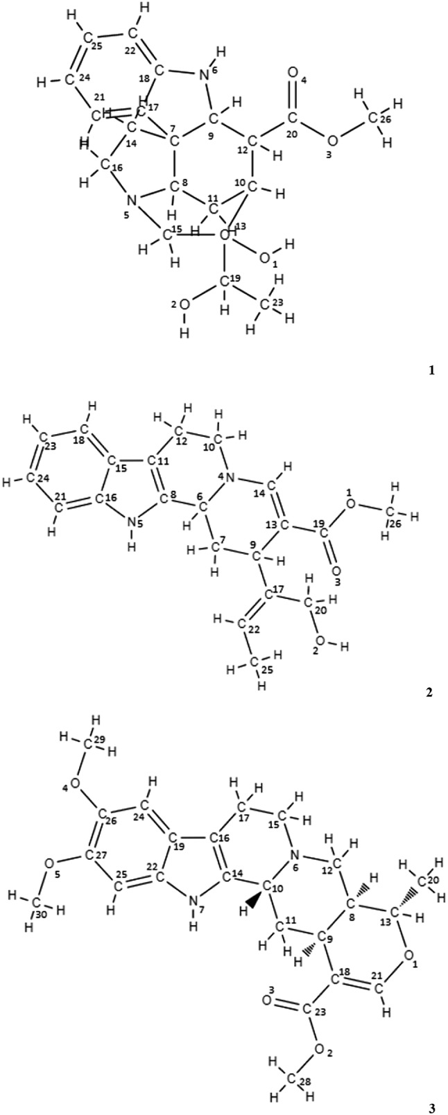
Using column chromatography and preparative TLC, three compounds were isolated from chloroform fraction of the leaf extracts of R. tetraphylla and the compounds showed positive result in Dragondroff’s test, Mayer’s test and Wagner’s test that confirmed the alkaloid nature of the compounds. The structures of the compounds were determined with the help of IR, GCMS and 1H and 13C NMR spectroscopy (Fig. 1).
Fig. 1.

Chemical structures of compounds 1–3 isolated from leaf extracts of R. tetraphylla
Compound 1 obtained as amorphous solid, and it showed retention time of 2.685 with 86.194% purity in HPLC. The mass spectrum analysis from GC–MS data showed compound with molecular weight 358 g/mol and Fragment ions m/z values 57, 144, 228, 358 that assigned to the molecular formula C20H26N2O4. IR absorptions at 3360 and 2930 cm−1 suggested the presence of alcohol and alkane (C–H). Three stretches at 1687, 1623 and 1437 cm−1 corresponds to double bonds and carboxylic acid group. The presence of amines and aromatic ring was confirmed by the stretches at 800 and 620 cm−1. The 1H NMR spectrum for compound 1 showed signals at δH 6.5–7.5, typical for an aromatic benzene ring system. The signals between δH 1.5 and 2.5 corresponding to allylic, benzylic or keto group in the compound. The signals obtained between δH 3.5 and 5.55 revealed the presence of alcohols and esters in the compound. The 13C NMR of the compound 1 showed 20 resonance spectrum values that are corresponding to carbon atoms attached with various bonding pattern of the structure. The signals obtained at δC 120.92, 122.5, 129.58, 149.89 and 172.03 showed carbon atoms present in the aromatic benzene ring structure, δC 44.64, 53.3 showed allylic, benzylic or keto group, δC 62.15, 61.71, 71.67 showed alcoholic and ester groups. The signals obtained from 1H and 13C NMR of compound 1 were compared with the signals of compound Curan-17-oic acid, 19, 20-dihydroxy, methyl ester (19S) and found resemblance in both GC–MS and NMR spectroscopy studies.
The molecular formula for compound 2 was established as C21H24N2O3 by GC–MS study that showed Fragment ions m/z values: 43, 57, 71, 149, 279 and 352 with molecular weight 352 g/mol. HPLC chromatogram for compound 2 showed retention time 2.160 with 73.026% purity. IR data for compound 2 showed various bonding pattern and functional groups such as 3390 cm−1 (Alcohol O–H), 1707 cm−1 (Carboxylic Acid C=O Stretch), 1210 cm−1 (Amine C–N Stretch), and 470 cm−1 (Aromatic C–H bend). The 1H NMR signals at δH 6.75, 6.92, 7.57, 7.1 and 7.4 corresponding to aromatic protons in benzene ring structure. The signals at δH 8.44 and 9.67 showed protons at heteroaromatic NH bonding system, δH at 1.94, 2.16, 2.35 showed protons at alkanes and keto groups, δH at 3.42, 3.54, 3.65, 3.68 showed protons at alcohol and ester groups, δH at 5.16 showed vinylic hydrogen atom bonded with alkene carbon atom. The 13C NMR spectrum data exhibited carbon atoms at δC 168, 155.5, 149.2, 144, 135.5, 126 and 120 corresponding to aromatic carbon atoms, δC signals at 115.7, 109.7, 106.6 showed carbon atoms in alcohol and ester groups, δC signals at 72.5, 59.9, 56.6, 53.5, 51.3, 38.6, 34.3, 31.5, 21.8 and 18.7 corresponding to carbon atoms at benzylic, keto, alkane groups. These evidences suggested that compound 2 was a yohimban class of alkaloid compound that was identified as 18, 19-Secoyohimban-19-oic acid, 16,17,20,21-tetrahydro-16-(hydroxymethyl)-methylester (Fig. 1) first time isolated from R. tetraphylla leaf extracts.
Compound 3 was isolated as amorphous solid and its molecular formula was established as C23H28N2O5 based on the Fragment ions m/z values 216, 283, 311, 397, 412 with molecular weight 412 g/mol in GC–MS. It showed sharp peak at retention time 2.167 with purity 83.436% in HPLC. IR spectrum for 3 exhibited various bonding pattern and functional groups such as 3407 cm−1 (Alcohol O–H and Amine N–H Stretch), 1089 cm−1 (Alcohol O–H stretch), 800 cm−1 (Amine N–H bend) and 480 cm−1 (Aromatic C–H stretch). IR spectra show numerous sharp bands between 500 and 1500 cm−1 which are assigned to deformation and stretching vibrations of the alkaloid ring system. The 1H NMR spectrum for compound 3 showed three signals at δH 6.32, 6.38 and 8.03 corresponding to hydrogen atom linked to aromatic carbon typically for a benzene ring. The δH signals at 3.54, 3.6 and 3.72 showed protons at three methoxy groups, δH signals at 1.18, 1.82, 1.96 showed protons at methylene groups. The 13C NMR spectrum data exhibited carbon signals at δC 134.37, 128.2, 136.83, 149.61, 148.97, 167.53, 154.63 corresponding to aromatic carbons present in a benzene ring structure. The δC values at 60.00, 56.67, 98.7, 93.9 and 73.63 showed carbon atoms in alcohol and ester groups, δC values at 56.13, 56.16 and 51.58 showed carbon atoms in allylic, benzylic and keto groups. Combined analysis of 1H and 13C NMR data of compound 3 indicated that the resonance values have resemblance with Reserpiline (Fig. 1) and it also confirmed by GC–MS analysis data.
In-vitro macrofilaricidal activity
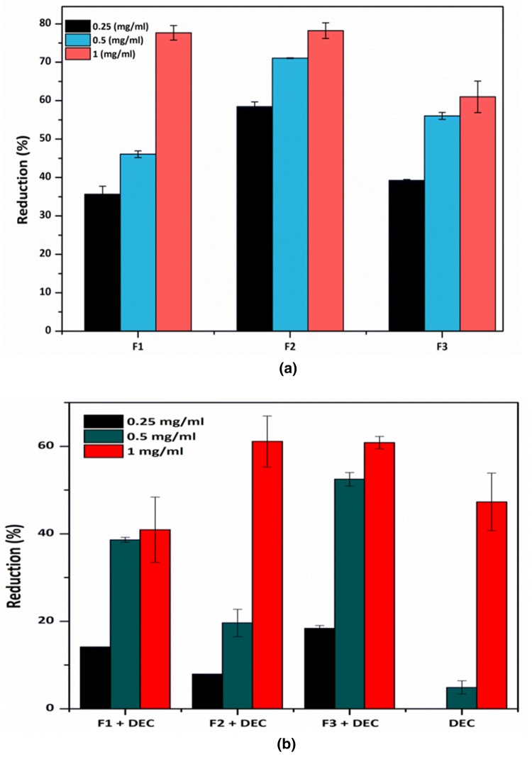
All compounds 1–3 showed significant adulticidal activities with IC50 values 0.54, 0.21 and 0.44 mg/ml (Fig. 2a) for compounds 1, 2 and 3 respectively in terms of MTT reduction assay that clearly proved the mortality of the adult worms as shown in Table 1 in different time intervals. DEC showed completely less active with percentage inhibition value of 47.31 ± 6.6 at 1.0 mg/ml (Fig. 2b). The combined effect of isolated compounds (F1, F2 and F3) and Diethylcarbamazine showed less effectiveness at the concentrations 0.25, 0.5 and 1.0 mg/ml than the compounds alone on Setaria worms. The combined effect was studied for assessing the effectiveness of compounds and DEC standard drug, as DEC alone is ineffective in killing adult parasites thus a combined effect may enhance the power of standard drug. But unfortunately DEC in combination with compounds 1–3 showed very low activity. The macrofilaricidal effect was further analysed by targeting the glutathione-S-transferase enzyme of S. cervi adult worms. This study showed that GST enzyme of adult parasites was significantly inhibited by the treatment of compounds 1–3 with percentage inhibition of 35.78%, 44.95% and 64.21% for compound 1, 2 and 3 respectively at concentration of 1.0 mg/ml (Table 2). The protein content estimation in each treated and control worm samples denoted their activity in vitro condition in a specific time.
Fig. 2.
a Macrofilaricidal effect of compounds 1–3 alone, b effects of compounds 1–3 in combination with DEC on adult parasites of S. cervi.
Notes: Vertical lines show mean ± SD. All the values obtained are statistically significant at p < 0.05 when compared with their respective controls. Parasites treated with DMSO were taken as control from which inhibition percentage was calculated
Table 1.
Effect of compounds 1–3 on the motility of adult parasites (S. cervi) in different time interval
| Compounds and DEC | Conc. (mg/ml) | Motility scores (in hours) | ||||
|---|---|---|---|---|---|---|
| 1 h | 2 h | 3 h | 4 h | 24 h | ||
| F1 | 0.25 | 4 + | 4+ | 4+ | 4+ | 2+ |
| F1 + DEC | 4+ | 4+ | 4+ | 4+ | 4+ | |
| F1 | 0.5 | 4+ | 4+ | 4+ | 4+ | 2+ |
| F1 + DEC | 4+ | 4+ | 4+ | 4+ | 4+ | |
| F1 | 1.0 | 4+ | 4+ | 4+ | 4+ | 1+ |
| F1 + DEC | 4+ | 4+ | 4+ | 4+ | 4+ | |
| F2 | 0.25 | 4+ | 4+ | 4+ | 4+ | 1+ |
| F2 + DEC | 4+ | 4+ | 4+ | 4+ | 4+ | |
| F2 | 0.5 | 4+ | 4+ | 4+ | 4+ | 1+ |
| F2 + DEC | 4+ | 4+ | 4+ | 4+ | 4+ | |
| F2 | 1.0 | 4+ | 4+ | 4+ | 4+ | 1+ |
| F2 + DEC | 4+ | 4+ | 4+ | 4+ | 3+ | |
| F3 | 0.25 | 4+ | 4+ | 4+ | 3+ | 1+ |
| F3 + DEC | 4+ | 4+ | 4+ | 4+ | 4+ | |
| F3 | 0.5 | 4+ | 3+ | 3+ | 2+ | 1+ |
| F3 + DEC | 4+ | 4+ | 4+ | 4+ | 4+ | |
| F3 | 1.0 | 4+ | 2+ | 1+ | 1+ | 1+ |
| F3 + DEC | 4+ | 4+ | 4+ | 4+ | 3+ | |
| DECa | 0.25 | 4+ | 4+ | 4+ | 4+ | 4+ |
| 0.5 | 4+ | 4+ | 4+ | 4+ | 4+ | |
| 1.0 | 4+ | 4+ | 4+ | 4+ | 4+ | |
All results are repeated three times for all groups
Motility scoring: 4+ (Highly motile), 3+ (Motile), 2+ (Sluggish), 1+ (non-motile) and Nil (Dead)
aStandard drug used as control
Table 2.
Effect of compounds 1–3 on the GST specific activity of adult parasite (S. cervi)
| Compounds | Conc. (mg/ml) | GST specific activity (µM/ml/min) | % Inhibition | Protein content (mg/ml) |
|---|---|---|---|---|
| F1 | 0.5 | 59.89 ± 2.3** | 15.6% | 2.13 ± 0.31* |
| 1.0 | 45.57 ± 1.8** | 35.78% | 2.39 ± 0.02* | |
| F2 | 0.5 | 59.24 ± 2.2** | 16.51% | 2.51 ± 0.51* |
| 1.0 | 39.06 ± 3.11** | 44.95% | 2.39 ± 0.06* | |
| F3 | 0.5 | 58.59 ± 0.88** | 17.43% | 2.46 ± 0.02* |
| 1.0 | 25.39 ± 1.32** | 64.21% | 2.06 ± 0.06* | |
| Control | – | 70.96 ± 1.21 | – | 1.67 ± 0.02 |
GST specific activity of S. cervi after treatment with compounds were compared with control and expressed as mean ± standard deviation (n = 3). Protein content was simultaneously estimated and also presented as mean ± standard deviation (n = 3). **Indicates p < 0.001 and *indicates p < 0.05 proved by one-way ANOVA followed by Dunnett’s two sided multiple comparison test as compared to control
In-silico molecular docking
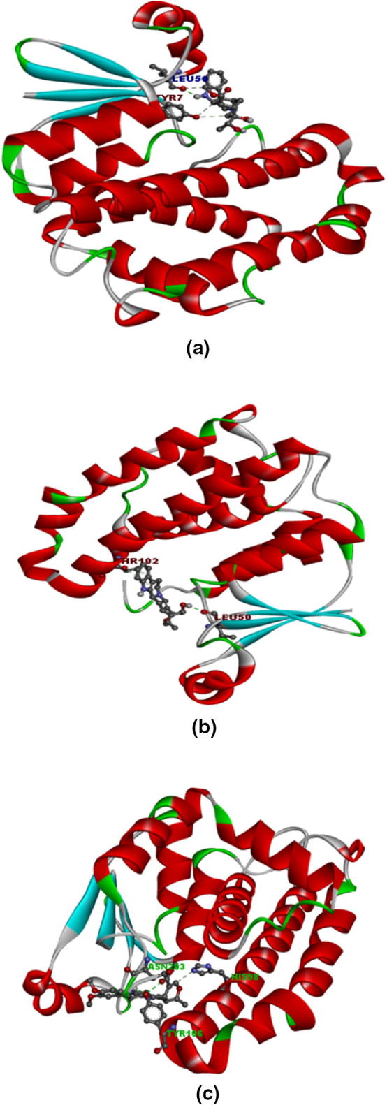
The present study revealed that Reserpiline with highest binding affinity of − 7.2 kcal/mol, second is the 18, 19-Secoyohimban with binding affinity of − 7.19 kcal/mol and then Curan-17-oic acid with binding affinity of − 5.14 kcal/mol (Table 3). These three compounds showed interaction by forming hydrogen bonds with amino acid residues of target GST molecule. The binding site amino acid residues that took part in bond formation are TYR-106, TYR-7, LEU-50, ASN-203, THR-102 (Fig. 3). This strong hydrophobic interaction may be the reason of high binding affinity, stability and activity of these isolated compounds.
Table 3.
Molecular docking analysis of ligands 1–3 with target GST enzyme (WbGST)
| Ligands | Binding energy (kcal/mol) | Ligand efficiency (kcal/mol) | Inhibition constant | Electrostatic energy | Hydrogen bond | Hydrophobic interaction |
|---|---|---|---|---|---|---|
| Curan-17-oic acid | − 5.14 | − 0.2 | 171.22 | 0.17 | LEU50, TYR7, TYR7, LEU50 | PHE8, PHE8 |
| 18, 19-Secoyohimban | − 7.19 | − 0.28 | 5.41 | − 0.11 | LEU50, THR102, THR102 | LEU13, LEU13, HIS98, LEU13 |
| Reserpiline | − 7.2 | − 0.24 | 5.25 | − 0.08 | TYR106, ASN203, TYR106, HIS98 | PHE8, PHE8, LEU13, LEU13, PHE8 |
Fig. 3.

Molecular docking study of three isolated compounds against target GST enzyme. a Compound 1 binding affinity with filarial GST, b compound 2 binding affinity with filarial GST and c compound 3 binding affinity with filarial GST. Stronger binding energy leads to better antifilarial efficacy of drugs
Discussion
Filariasis imparts a huge socio-economic burden in tropical and sub-tropical countries. According to WHO, Filariasis is one of the debilitating vector borne disease second to Malaria in Asian countries. The conventional drugs such as DEC, albendazole and ivermectin are more microfilaricidal than macrofilaricidal and also possess side effects. The development of a natural safer drug is therefore urgently needed that would surely effective against adult parasites of filarial disease. In this regard, a promising ethno-medicinal plant R. tetraphylla was explored for effective antifilaricidal principles and computational study was undertaken for the evaluation of affinity of isolated compounds with antifilarial drug target i.e. filarial GST enzyme.
In this study, isolation of active principles resulted into three compounds namely, Compound F1 i.e., Curan-17-oic acid, 19, 20-dihydroxy, methyl ester (19s) has been reported first time from R. tetraphylla plant leaf extract. Previous reports on isolation and characterization of compounds by gas chromatography and mass spectroscopy analysis of methanolic leaves extract of Adiantum capillus-veneris revealed Curan-17-oic acid compound that shows antimicrobial and anti-inflammatory activities (Hussein et al. 2016). GC–MS analysis of methanolic extract of Eucalyptus citriodora also exhibited Curan-17-oic acid and bioactivities such as antidiabetic and insecticidal properties reported from for that compound (Sahi 2016). Compound F2 i.e., 18, 19-seco-yohimban has also been reported from plants such as Sesamum radiatum essential oil of the leaves, methanol extract of Eichhornia Crassipes (waterhyacinth), methanol extract of Basella alba leaf (Ogunlesi et al. 2010; Shanab et al. 2010; Baskaran et al. 2015). Yohimbine has been found active on endocrine and reproductive systems. Yohimbine has also been found to exhibit cardiovascular activity (Henauer et al. 1984). Compound F3 identified as Reserpiline i.e., one of the well known alkaloid earlier reported from root bark and stem bark of Rauvolfia vomitoria, roots of Rauvolfia serpentina and from chloroform and methanol extract of leaves of R. tetraphylla (Poisson 1959; Srivastava et al. 2006; Maurya et al. 2013). Reserpiline has antipsychotic, antigastric secretion, antihypertension activities. In the present study this compound has studied first time for antifilarial property on S. cervi.
The in silico molecular docking study was performed to determine the binding affinity of three isolated compounds (F1, F2 and F3) with the target enzyme pi-class GST of W. bancrofti. Earlier studies on GST enzyme of W. bancrofti revealed that WbGST shares 98% identity with B. malayi GST, 79% identity with O. volvulus GST and 75% identity with D. immitis GST. This report has confirmed by multiple sequence alignment of amino acid sequences of WbGST with other filarial nematode GSTs (Veerapathran et al. 2009). Thus to enumerate the interaction mechanism of pi-class glutathione-S-transferase (detoxification enzyme) of W. bancrofti (nematode parasite) with the selected significant ligands from the plant R. tetraphylla, molecular docking was undertaken We have also attempted the same for our isolated molecules and results are promising.
Previous reports on docking of 58 phytochemicals with nematicidal, anthelmintic and GST substrate activities shows that only Curcumin, Vanillin and Brucine have highest binding affinity of − 8.4343, − 7.0792 and − 4.2539 kcal/mol respectively with GST enzyme (Azeez et al. 2012). It has also been reported that the molecular docking of reference drugs DEC and Ivermectin with nematode GST enzyme reveals the binding energy as − 4.9 kcal/mol and − 8.4 kcal/mol respectively (Kalani et al. 2014). This study also stated about the toxicity of these reference drugs by examining through MetaDrug tool (Thomson Reuters, USA).
Conclusions
The present study has provided leads from R. tetraphylla leaves for macrofilaricidal drug development against filariasis. To our knowledge, this study is the first to demonstrate the macrofilaricidal activity of three compounds namely, Curan-17-oic acid, 19, 20-dihydroxy, methyl ester (compound 1), 18, 19-Secoyohimban-19-oic acid (compound 2) and Reserpiline (compound 3) isolated from leaves of R. tetraphylla. The result produced in this study might be additional reference in natural product research and will contribute to the further study of antifilarial drug discovery.
Acknowledgements
The authors want to thank the Forest and Environment Dept., Govt. of Odisha for providing funding support in the form of state plan project for smooth completion of research work.
Author’s contributions
DRB contributed in collecting plant samples and parasite, isolated the compounds and performed all in vitro and in silico tests on parasites. SB contributed in data interpretation and critical reading of the manuscript.
Compliance with ethical standards
Conflict of interest
No potential conflict of interest was reported by the authors.
References
- Ahmad R, Srivastava AK. Inhibition of filarial glutathione S-transferase by various classes of compounds and their evaluation as novel antifilarial agents. Helminthologia. 2008;45(3):114–120. doi: 10.2478/s11687-008-0022-3. [DOI] [Google Scholar]
- Ardelli BF, Guerriero SB, Prichard RK. Genomic organization and effects of ivermectin selection on Onchocerca volvulus P-glycoprotein. Mol Biochem Parasitol. 2005;143:58–66. doi: 10.1016/j.molbiopara.2005.05.006. [DOI] [PubMed] [Google Scholar]
- Azeez S, Babu RO, Aykkal R, Narayanan R. Virtual screening and in vitro assay of potential drug like inhibitors from spices against glutathione-S-transferase of filarial nematodes. J Mol Model. 2012;18:151–163. doi: 10.1007/s00894-011-1035-2. [DOI] [PubMed] [Google Scholar]
- Baskaran G, Salvamani S, Ahmad SA, Shaharuddin NA, Pattiram PD, Shukor MY. HMG-CoA reductase inhibitory activity and phytocomponent investigation of Basella alba leaf extract as a treatment for hypercholesterolemia. Drug Deg Dev Therapy. 2015;9:509–517. doi: 10.2147/DDDT.S75056. [DOI] [PMC free article] [PubMed] [Google Scholar]
- Behera DR, Bhatnagar S. Macrofilaricidal activity of leaf extracts of Rauvolfia tetraphylla L. against bovine filarial parasite Setaria cervi. Int J Pharm Phytochem Res. 2017;9(9):1217–1222. [Google Scholar]
- Brophy PM, Pritchard DI. Parasitic helminth glutathione S-transferases: an update on their potential as targets for immunoand chemotherapy. Exp Parasitol. 1994;79:89–96. doi: 10.1006/expr.1994.1067. [DOI] [PubMed] [Google Scholar]
- Campbell AM, van Eldik AJ, Liebau E, Barrett J, Brophy PM, Teesdale-Spittle PH, Wang MF. Towards validation of glutathione S-transferase (GST) as a filarial nematode drug target. Chem Biol Interact. 2001;133:240–243. [Google Scholar]
- Comley JCW, Rees MJ, Turner CH, Jenkins DC. Colorimetric quantitation of filarial viability. Int J Parasitol. 1989;19(1):77–83. doi: 10.1016/0020-7519(89)90024-6. [DOI] [PubMed] [Google Scholar]
- Dreyer G, Noroes J, Addiss D. The silent burden of sexual disability associated with lymphatic filariasis. Acta Trop. 1997;63(1):57–60. doi: 10.1016/S0001-706X(96)00604-3. [DOI] [PubMed] [Google Scholar]
- Habig WH, Pabst MJ, Jakoby WB. Glutathione S-transferases the first enzymatic step in mercapturic acid formation. J Biol Chem. 1974;249:7130–7139. [PubMed] [Google Scholar]
- Henauer SA, Gillespie HK, Hollister LE. Yohimbine and the model anxiety state. J Clin Psychiatry. 1984;45:512–515. [PubMed] [Google Scholar]
- Hussein HM, Hameed IH, Ibraheem OA. Antimicrobial Activity and spectral chemical analysis of methanolic leaves extract of Adiantum Capillus-Veneris using GC-MS and FTIR spectroscopy. Int J Pharm Phytochem Res. 2016;8(3):369–385. [Google Scholar]
- Kalani K, Kushwaha V, Sharma P, Verma R, Srivastava M. In Vitro, in silico and in vivo studies of ursolic acid as an anti-filarial agent. PLoS ONE. 2014;9(11):e111244. doi: 10.1371/journal.pone.0111244. [DOI] [PMC free article] [PubMed] [Google Scholar]
- Kardono LBS, Tsauri S, Padmawinata K, Pezzuto JM, Kinghorn AD. Cytotoxic constituents of the bark of Plumeria rubra collected in Indonesia. J Nat Prod. 1990;53(6):1447–1455. doi: 10.1021/np50072a008. [DOI] [PubMed] [Google Scholar]
- Kaushal NA, Kaushal DC, Ghatak S. Identification of antigenic proteins of Setaria cervi by immunoblotting technique. Immun Invest. 1987;16:139–149. doi: 10.3109/08820138709030571. [DOI] [PubMed] [Google Scholar]
- Lakshmi V, Joseph SK, Srivastava S, Verma SK, Sahoo MK, et al. Antifilarial activity in vitro and in vivo of some flavonoids tested against Brugia malayi. Acta Trop. 2010;116:127–133. doi: 10.1016/j.actatropica.2010.06.006. [DOI] [PubMed] [Google Scholar]
- Lazdins J, Kron M. New molecular targets for filariasis drug discovery. Parasitol Today. 1999;15(8):305–306. doi: 10.1016/S0169-4758(99)01483-0. [DOI] [PubMed] [Google Scholar]
- Lowry OH, Rosebrough NJ, Farr AL, Randall RJ. Protein measurement with the Folin phenol reagent. J Biol Chem. 1951;193:265–276. [PubMed] [Google Scholar]
- Malinowska I, Gadzikowska M, Waksmundzka-Hajnos M. Mobile-phase velocity: a tool for separation of alkaloids by OPLC. J Planar Chromatogr. 2005;18:176. doi: 10.1556/JPC.18.2005.3.1. [DOI] [Google Scholar]
- Masoko P, Eloff JN. Screening of twenty-four south african combretum and six Terminalia species (combretaceae) for antioxidant activities. Afr J Tradit CAM. 2007;4:231–239. doi: 10.4314/ajtcam.v4i2.31213. [DOI] [PMC free article] [PubMed] [Google Scholar]
- Mathew N, Paily NKP, Vanamail AP, Kalyanasundaram M, Balaraman K. Macrofilaricidal activity of the plant Plumbago indica/rosea in vitro. Drug Dev Res. 2002;56:33–39. doi: 10.1002/ddr.10056. [DOI] [Google Scholar]
- Maurya A, Gupta S, Srivastava SK. Large-scale separation of antipsychotic alkaloids from Rauwolfia tetraphylla L. by pH-zone-refining fast centrifugal partition chromatography. J Sep Sci. 2013;36:407–413. doi: 10.1002/jssc.201200273. [DOI] [PubMed] [Google Scholar]
- Mukherjee M, Misra S, Chatterjee RK. Development of in vitro screening system for assessment of antifilarial activity of compounds. Acta Trop. 1998;70:251–255. doi: 10.1016/S0001-706X(98)00016-3. [DOI] [PubMed] [Google Scholar]
- Murthy PK. Evaluation of two in vitro systems employing Brugia malayi parasite for prescreening of potential antifilarials. Curr Sci. 1999;77(8):1084–1089. [Google Scholar]
- Ogunlesi M, Okiei W, Osibote EA. Analysis of the essential oil from the leaves of Sesamum radiatum, a potential medication for male infertility factor, by gas chromatography–mass spectrometry. Afr J Biotech. 2010;9(7):1060–1067. doi: 10.5897/AJB09.941. [DOI] [Google Scholar]
- Panwar GS, Guru SK. Alkaloid profiling and estimation of Reserpine in Rauwolfia serpentina plant by TLC, HPTLC and HPLC. Asian J Plant Sci. 2011;10(8):393–400. doi: 10.3923/ajps.2011.393.400. [DOI] [Google Scholar]
- Poisson J. Research on the alkaloids of Rauwolfia vomitoria Afz. roots (Apocynaceae) Trav Lab Matiere Med Pharm Galenique Fac Pharm Paris. 1959;44:2–118. [PubMed] [Google Scholar]
- Rajesh A, Shamsudin M. In silico molecular docking studies on phytocompounds from the plant Kalanchoe pinnata targeting the pi-class glutathione-s-transferase of Wuchereria bancrofti. Int J Zool Appl Biol. 2017;2(5):258–265. [Google Scholar]
- Sahare KN, Singh V. Antifilarial activity of ethyl acetate extract of Vitex negundo leaves in vitro. Asian Pac J Trop Med. 2013;6:689–692. doi: 10.1016/S1995-7645(13)60119-4. [DOI] [PubMed] [Google Scholar]
- Sahi NM. Evaluation of insecticidal activity of bioactive compounds from Eucalyptus citriodora against Tribolium castaneum. Int J Pharm Phytochem Res. 2016;8(8):1256–1270. [Google Scholar]
- Schwab AE, Boakye DA, Kyelem D, Prichard RK. Detection of benzimidazole resistance-associated mutations in the filarial nematode Wuchereria bancrofti and evidence for selection by albendazole and ivermectin combination treatment. Am J Trop Med Hyg. 2005;73:234–238. doi: 10.4269/ajtmh.2005.73.234. [DOI] [PubMed] [Google Scholar]
- Shanab SMM, Shalaby EA, Lightfoot DA, El-Shemy HA. Allelopathic effects of water hyacinth [Eichhornia crassipes] Plus One. 2010;5:e13200. doi: 10.1371/journal.pone.0013200. [DOI] [PMC free article] [PubMed] [Google Scholar]
- Sofowora A. Medicinal plants and traditional medicines in Africa. Ibadan: Spectrum Books; 1993. p. 150. [Google Scholar]
- Srinivasan L, Mathew N, Karunan T, Muthswamy K. Biochemical studies on glutathione S-transferase from the bovine filarial worm Setaria digitata. Parasitol Res. 2011;109:213–219. doi: 10.1007/s00436-010-2227-x. [DOI] [PubMed] [Google Scholar]
- Srivastava A, Tripathi AK, Pandey R, Verma RK, Gupta MM. Quantitative determination of reserpine, ajmaline and ajmalicine in Rauvolfia serpentina by reversed-phase high-performance liquid chromatography. J Chromatogr Sci. 2006;44:557–560. doi: 10.1093/chromsci/44.9.557. [DOI] [PubMed] [Google Scholar]
- Trease GE, Evans WC. Pharmacognosy. 13. London: Bailliere Tindall; 1989. pp. 176–180. [Google Scholar]
- Veerapathran A, Dakshinamoorthy G, Gnanasekar M, Reddy MVR, Kalyanasundaram R. Evaluation of Wuchereria bancrofti GST as a vaccine candidate for lymphatic filariasis. PLoS Negl Trop Dis. 2009;3(6):e457. doi: 10.1371/journal.pntd.0000457. [DOI] [PMC free article] [PubMed] [Google Scholar]
- WHO Global programme to eliminate lymphatic filariasis. Wkly Epidemiol Record. 2009;84:437–444. [PubMed] [Google Scholar]


