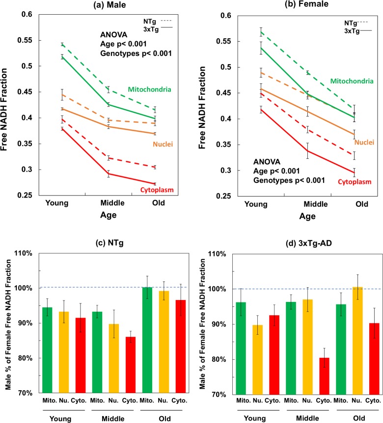
Fig. 3.
Mitochondrial, nuclear, and cytoplasmic free NADH fractions decline with age are further depleted with AD genotype of both a male and b female mouse neurons. For the compartment effect, mitochondria were the most reduced with the highest free NADH fractions and cytoplasm presents the most oxidized NADH state with the lowest free NADH levels (ANOVA for the compartment-differences, in male, NTg F(2,89) = 397, p < 0.001, 3xTg-AD F(2, 89) = 927, p < 0.001; in female, NTg F(2,179) = 154, p < 0.001, 3xTg-AD F(2,179) = 106, p < 0.001). Aging depleted free NADH levels in all compartments of both genotypes. ANOVA for male, mitochondria F(2,59) = 338, p < 0.001, nuclei F(2,59) = 56, p < 0.001, cytoplasm F(2,59) = 262, p < 0.001; for female, mitochondria F(2,119) = 161, p < 0.001, nuclei F(2,119) = 51, p < 0.001, and cytoplasm F(2,119) = 96, p < 0.001). The 3xTg-AD genotype demonstrated significantly more bound NADH and more oxidized NADH redox state than the age-matched NTg neurons in each compartment. By gender (c, d), male neurons were lower in free NADH fraction than the female neurons at young and middle ages, but the multiple comparison results indicated no significant differences in old ages between female and male neurons. Two-way ANOVA for gender effect on compartments, mitochondria F(1,59) = 13, p < 0.001; nuclei F(1,59) = 13, p < 0.001; cytoplasm F(1,59) = 25, p < 0.001). In 3xTg-AD neurons, male NADH fraction was lower than that in female neuron of each compartment (mitochondria F(1,59) = 7, p < 0.01; nuclei F(1,59) = 8, p < 0.01; cytoplasm F(1,59) = 51, p < 0.001) and each age (young F(1,59) = 18, p < 0.001; middle F(1,59) = 26, p < 0.001; old F(1,59) = 8, p < 0.01)

