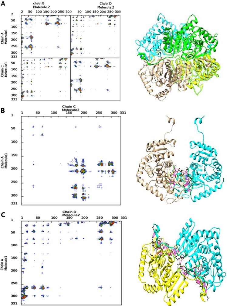
Figure 1.
contact map and 3D structure of dimers and tetramer form of the enzyme. 3D structures of LDHA is depicted in the right as tetramer form (A), (A–C) dimer form (B), and (A–D) dimer form (C). In the left, the contact maps of the intermolecular contacts have been depicted as the colored dots. Red, yellow, green, and blue indicate contacts within 7 Å, 10 Å, 13 Å and 16 Å, respectively. Also violet displays hydrophilic-hydrophilic, green shows hydrophobic-hydrophobic and yellow shows hydrophilic-hydrophobic interactions.

