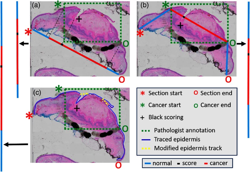
Fig. 2.
Methods for the mapping of cancer annotations to the “annotation line” of fixed tissue bread-loaf blocks. The methods include (a) line projection, (b) trapezoidal projection, and (c) epidermis-extraction projection. The H&E slide is shown with the pathologists’ annotations. Section start and end points, cancer start and end points, and black scoring point are identified accordingly. The final lines to superimpose on the epidermal image are shown as vertical lines on the sides that are colored red (cancer), blue (noncancer), and black (black scoring point). For the epidermis-extraction projection, the curve is manually smoothened to avoid the extra distance due to tears and debris. See previous text for additional details.

