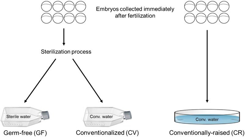
Fig. 1.
Schematic illustrating the development of gnotobiotic zebrafish groups. Immediately after fertilization, zebrafish embryos were collected and randomly split into 3 groups. One group was collected directly into a sterile petri dish containing conventional fish water (CR group). The other 2 groups of embryos were sterilized and then collected into tissue culture flasks containing either sterile fish water (GF group) or conventional fish water (CV group). Embryos were maintained at ~1 embryo/mL until testing at 6 dpf.

