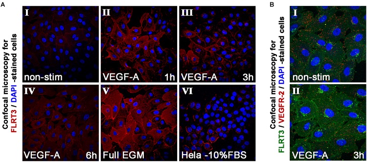
FIGURE 4.
Immunofluorescent staining of FLRT3. (A) Immunofluorescent staining of FLRT3 confirmed VEGF-A-induced upregulation of FLRT3 also at the protein level and its internalization and localization from cell surface into cytoplasm and small intracellular vesicles near the nucleus (panels I–IV, representative pictures of non-stimulated and VEGF-A-stimulated HUVECs at 1–6 h time points). A part of the positivity for FLRT3 was retained also at cell surface, especially on areas where adjacent HUVECs were in contact to each other (panels II–III). Higher expression of FLRT3 was detected in proliferating HUVECs grown in high serum conditions (V). Hela cells expressing low quantity of endogenous FLRT3 were used as negative controls for the immunofluorescent stainings (VI). (B) Immunofluorescent double-staining for FLRT3 and VEGFR-2 in non-stimulated and VEGF-A-stimulated HUVECs 3 h post-treatment.

