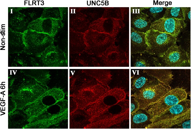
FIGURE 5.
Immunofluorescent double-staining for FLRT3 and UNC5B in non-stimulated (I–III) and VEGF-A-stimulated (IV–VI) HUVECs 6 h post-treatment. Co-localization of these factors was detected especially in VEGF-A-stimulated HUVECs and took place on cell surface as well as intracellular vesicles near the nucleus (VI). Immunofluorescent stainings for FLRT3 (I, IV), UNC5B (II, V), and merged images including nuclear staining (blue color; III, VI).

