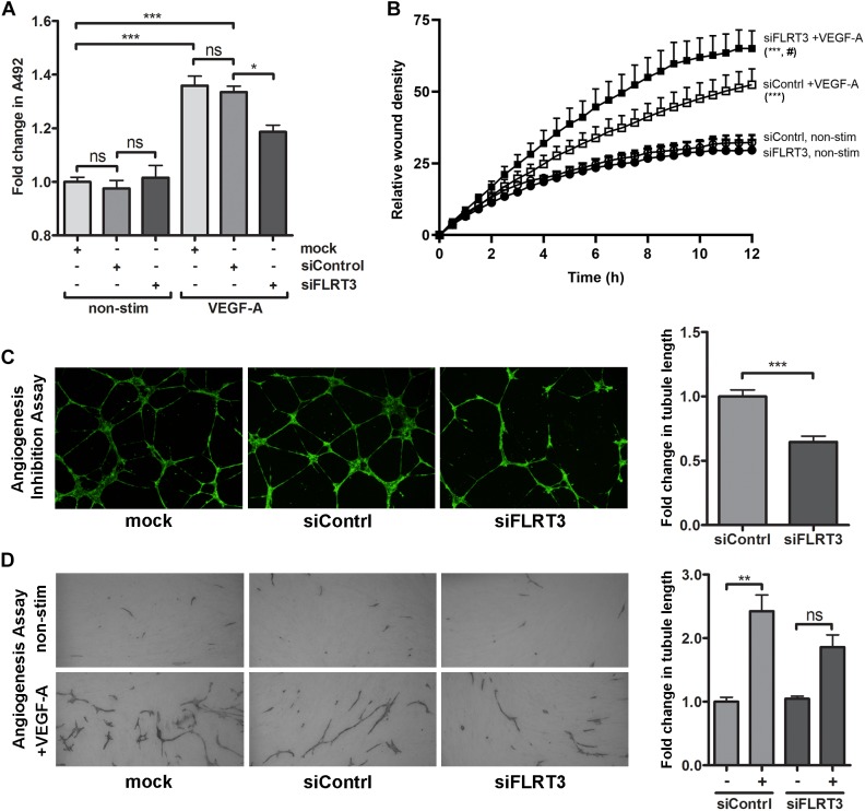
FIGURE 9.
Transcriptional inhibition of FLRT3 in HUVECs decreased VEGF-A-stimulated cell survival and in vitro angiogenesis but enhanced cell migration. (A) In MTS assay, the ability of VEGF-A (50 ng/ml) to stimulate EC survival was significantly decreased in the HUVECs transfected with siRNA against FLRT3. (B) VEGF-A-stimulated cell migration, assessed as a relative wound density (RWD), was significantly enhanced over the time in HUVECs transfected with FLRT3 siRNA as compared to control siRNA-treated control cell. Non-stimulated HUVECs for both study groups was included to see the background level of RWD in each time point and cellular migration was monitored continuously using the IncuCyte S3 Live-cell Imaging System. (C,D) FLRT3 siRNA significantly decreased tube formation in in vitro angiogenesis models. The data presented as mean ± SEM are representative of repeated independent experiments done in 3–8 replicates. For the experiments in (A,C,D) ∗p < 0.05; ∗∗p < 0.01; ∗∗∗p < 0.001; #p < 0.01 as compared to corresponding cells transfected with control siRNA; and ns, non-significant. (B) Statistical significance between the study groups was quantified by comparing the main column effects over the time. ∗∗∗, #p < 0.001 as compared to corresponding non-treated cells or to the VEGF-A-stimulated cells transfected with control siRNA, respectively.

