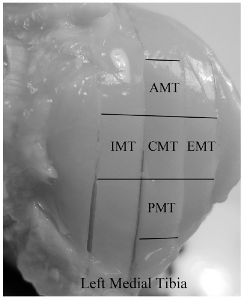
Figure 1.

The topographical locations of the specimens on the surface of a left medial tibia. In the meniscus-covered area, there are anterior medial tibia (AMT), exterior medial tibia (EMT) and posterior medial tibia (PMT) specimens. In the meniscus-uncovered area, there are central medial tibia (CMT) and interior medial tibia (IMT) specimens.
