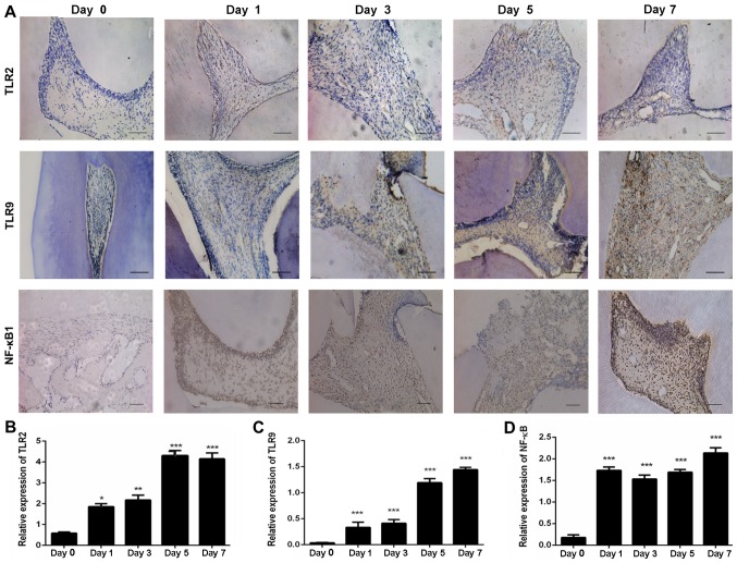
Figure 2.
Immunochemical staining for TLR2, TLR9 and NF-κB1 in pulp tissue sections. (A) Images of staining for TLR2, TLR9 and NF-κB1 expression in rat IP tissues (scale bar, 100 µm). (B) TLR2, (C) TLR9 and (D) NF-κB1 expression levels were significantly upregulated in the IP group on days 1–7 as compared with the day 0 (control) group. The experiments were repeated in triplicate, and the results were analyzed using ImageJ software. Error bars represent the standard error of the mean. *P<0.05, **P<0.01 and ***P<0.001 vs. day 0. TLR, toll-like receptors; NF-κB1, nuclear factor-κB1; IP, irreversible pulpitis.

