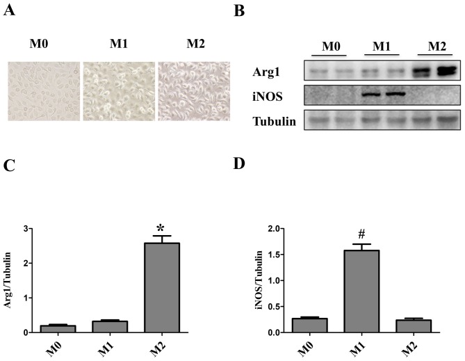
Figure 1.
Identification of macrophages. Bone marrow-derived macrophages (M0) were treated for 24 h with lipopolysaccharide (100 ng/ml) + interferon-γ (6 ng/ml) to induce M1 polarization or IL-4 (10 ng/ml) + IL-13 (10 ng/ml) to induce M2 polarization. (A) Macrophage morphology was observed under phase contrast microscopy (magnification, ×400): M0 macrophages were elongated and spindle-shaped, M1 macrophages were round and M2 macrophages were cone-shaped. (B) Western blot analysis was performed to determine the protein expression levels of iNOS, an M1 marker and Arg1, an M2 marker. Tubulin was used as the loading control. (C and D) Quantification of the western blot results. The data are presented as the mean ± standard error of the mean. *P<0.05 vs. M0; #P<0.05 vs. M0. Arg1, arginase1; iNOS, inducible nitric oxide synthase; M, macrophage.

