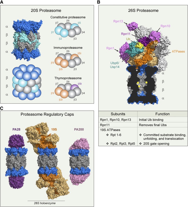
Fig. 2.
Proteasome structure and function. (A) Structures (PDB 4r3o) and cartoon representation of 20S proteasome, highlighting the different β-subunit combinations found in tissue-specific proteasomes discussed in the text. (B) Structure of the 26S proteasome in complex with Ubp6 (PDB 5a5b). A cross-section of 20S proteasome reveals the C terminus of Rpt5 ATPase (dark orange) positioned in the inter-α-subunit pocket (asterisk). Proteolytic sites are marked with yellow stars. Labeled 19S subunits are discussed in the text. (C) 20S proteasomes (blue and gray) complexed with regulatory caps: PA28 homolog PA26 (PDB 1fnt), 19S (PDB 5gjr), and PA200 yeast homolog Blm10 (PDB 4v7o). 19S ATPases are dark orange, and non-ATPase subunits are light orange. PDB, Protein Data Bank.

