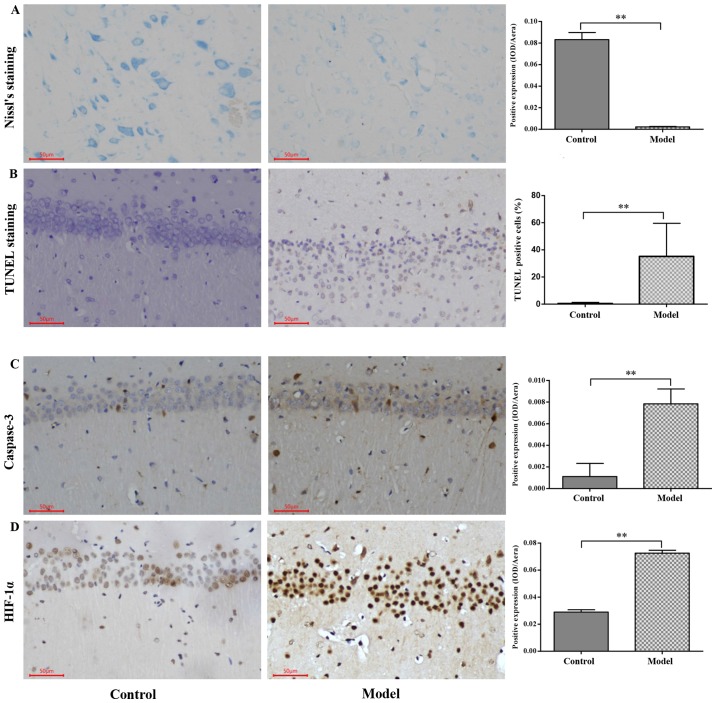
Figure 4.
Immunohistochemistry for caspase-3 and HIF-1α protein expression and Nissl's and TUNEL staining in the CA1 region in the hippocampus following hypoxia. (A) Nissl-stained neurons in the CA1 region of the hippocampus of SD rats. (B) TUNEL-positive apoptotic cells in the CA1 region of the hippocampus of SD rats. (C) Caspase-3 expression examined using immunohistochemical analysis and quantified. (D) HIF-1α expression examined using immunohistochemical analysis and quantified. Scale bar, 50 µm. Original magnification, ×200. The values are presented as the mean ± standard deviation. **P<0.01 with comparisons shown by lines. HIF-1α, hypoxia inducible factor-1α; TUNEL, terminal deoxynucleotidyl transferase dUTP nick end labeling; SD, Sprague-Dawley; IOD, integrated optical density.

