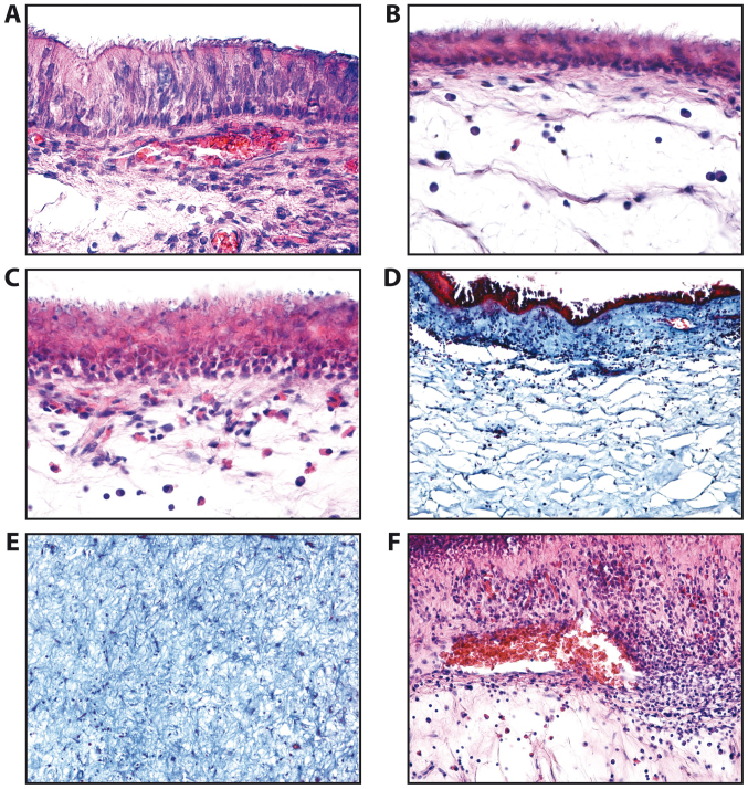
Figure 1.
Epithelial types observed in the nasal mucosa and polyps of patients with chronic rhinosinusitis. (A) Normal epithelium (H&E staining, ×200 magnification). (B) Atrophic epithelium squamous metaplasia of epithelium (H&E staining, ×200 magnification). (C) Squamous metaplasia of the epithelium (H&E staining, ×200 magnification). (D) Lamina propria of nasal polyp (H&E staining, ×100 magnification). (E) Fibrous areas of lamina propria of nasal polyp (H&E staining, ×100 magnification). (F) Nasal polyp; areas with vascular congestion (H&E staining, ×100 magnification).

