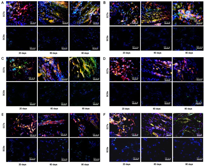
Figure 7.
Immunostaining for connexin-40, connexin-43, HCN2, connexin-45, HCN4 and cTnT in the transplanted ECTs (n=26) and BCSs (n=24). Representative staining for (A) connexin-40, (B) connexin-43, (C) HCN2, (D) connexin-45, (E) HCN4 and (F) cTnT in the rats transplanted with ECTs and BCSs at days 20, 60, and 90 after implantation. The transplanted tissue cells were identified by labeling with CM-Dil (red). Immunostaining for connexin-40, connexin-43, HCN2, connexin-45, HCN4 and cTnT in the implantation site was shown by the secondary antibodies (green). The nuclei were counterstained with DAPI (blue). ECTs, engineered conduction tissues; BCSs, blank collagen sponges.

