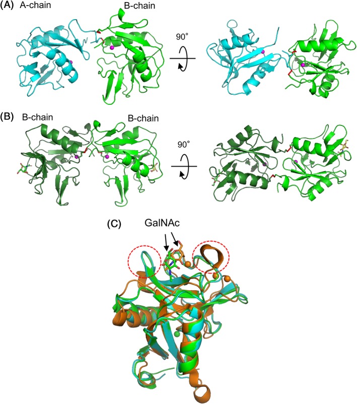
Figure 6.

Overall structures of SPLs. (A) SPL‐1 composed of A‐chain (cyan) and B‐chain (green) (PDB ID: 6A7T). (B) SPL‐2/GalNAc complex composed of two B‐chains (PDB ID: 6A7S). (C) A‐ and B‐chains of SPLs and a subunit of C. echinata C‐type lectin, CEL‐I (PDB ID: 1WMZ) complexed with GalNAc,11 are superposed based on their main‐chain structure using PyMOL.40 Bound GalNAc molecules are depicted as stick models. Magenta spheres indicate bound Ca2+ ions.
