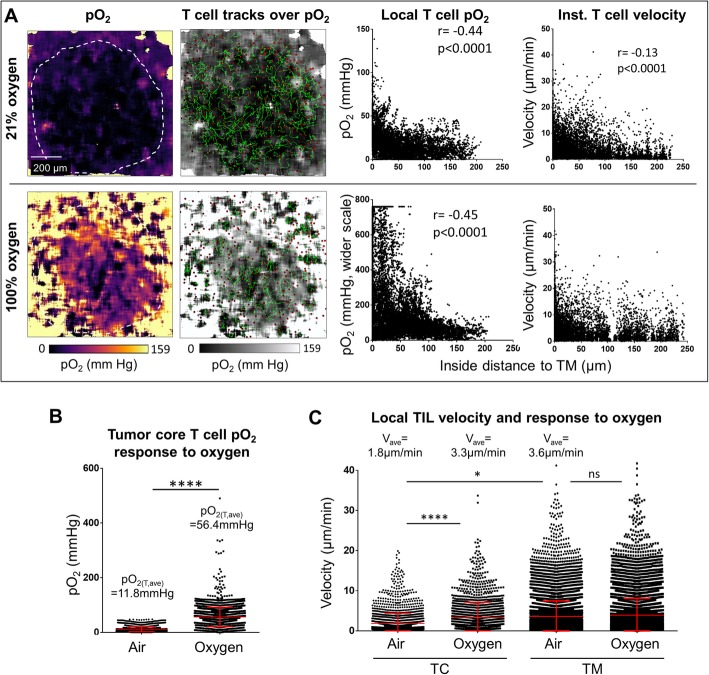
Fig. 4.
Response of tumor infiltrating T cell motility to changing the specimen breathing from air (21% oxygen) to 100% oxygen. a The same tumor as in Fig. 3 was imaged by FaST-PLIM while the mouse was breathing air (upper row) or about 30 min after breathing change to oxygen (lower row). From the left: images of oxygen tensions (pO2), T cell tracks overlaid on pO2; relation of intratumoral T cell pO2 experience to the distance to tumor margin; corresponding relation of T cell instantaneous velocity to the distance to tumor margin. b Local pO2 for T cells located in tumor core when the mouse was breathing air vs. that for breathing oxygen. c Local velocities of tumor infiltrating T cells (TIL) in tumor core (TC) or tumor margin (TM) while the mouse was breathing air and after breathing oxygen. Each symbol represents a T cell instance (n > 5000). Graphs display data from representative tumor FOV. * denotes p < 0.05 determined by Student’s T-test. Vertical bars represent standard deviations. r = Pearson’s coefficient

