Fig. 2.
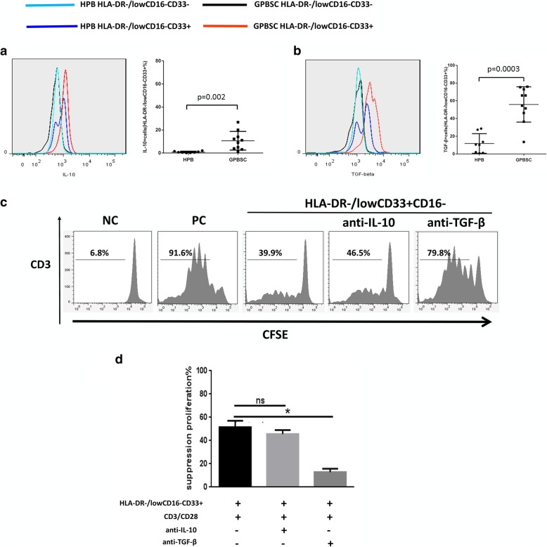
The percentage of IL-10+ and TGF-β+ cells increased in HLA-DR−/lowCD33+CD16− cells in GPBSC and HLA-DR−/lowCD33+CD16− cells suppressed T cell proliferation in a TGF-β-dependent manner. a, b IL-10 and TGF-β were analyzed by cytometry in HPB and GPBSC from eight healthy donors. IL-10+ and TGF-β+ cells were gated on HLA-DR−/lowCD33+CD16−cells. c HLA-DR−/lowCD33+CD16− cells sorted from GPBSC and co-cultured with autologous CD3+ T cells, with or without anti-IL-10 antibody or anti-TGF-β antibody in culture system in the presence of anti-CD3/CD28 beads. The ratio of HLA-DR−/lowCD33+CD16− to CD3+ T cells was 1:1. Co-cultured for 4 days, T cell proliferation was evaluated using CFSE labeling. Unstimulated T cells were used as a negative control. Stimulated T cells were used as a positive control. d The percentage of T cell suppression was shown in a different group. Data were compared using Student’s unpaired t test (ns, not significant; *p ≤ 0.05). d Percentages of T cell suppression in different groups. Data were compared using Student’s unpaired t test (ns, not significant; *p ≤ 0.05)

