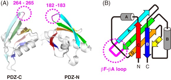
Figure 1.

3D structure of the PDZ tandem from the A. aeolicus Site‐2 protease homolog. (A) Ribbon model of PDZ tandem. The six β‐strands of the respective PDZ domains are colored differently where PDZ‐N and PDZ‐C are shown in bright and pale colors, respectively. The deleted loop residues for the PA‐insertion in the β‐hairpins are indicated in magenta with dotted circles. (B) Topology diagram of the circular‐permutant PDZ domain. The loop connecting the βF and βA strands is colored in magenta and indicated with a dotted circle. In the present work, the PA tag was inserted here.
