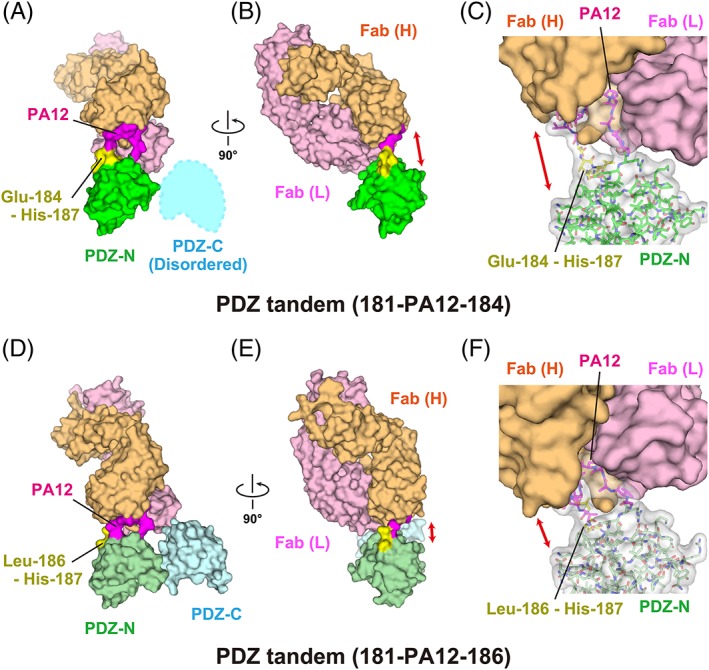
Figure 2.

Complex formation of the PDZ tandem with the NZ‐1 Fab through a PA‐inserted PDZ‐N domain. (A, B) Surface model of PDZ tandem (181‐PA12‐184) in complex with the NZ‐1 Fab in two different views. The inserted PA tag is shown in magenta. The residues undergoing significant structural changes compared with the wild type, as shown in Figure 5, are highlighted in yellow. (C) Close‐up view of the binding site. The PDZ‐N domain and the inserted PA tag are shown as stick models with a transparent surface. The solvent‐accessible space between the rigidly folded part of the PDZ‐N domain and the NZ‐1 Fab is indicated with a double‐headed arrow. (D, E) Surface model of PDZ tandem (181‐PA12‐186) in complex with the NZ‐1 Fab in two different views. (F) Close‐up view of the binding site. The models are colored as in (A, B, and C) while the PDZ domains are shown in pale colors.
