Figure 1.
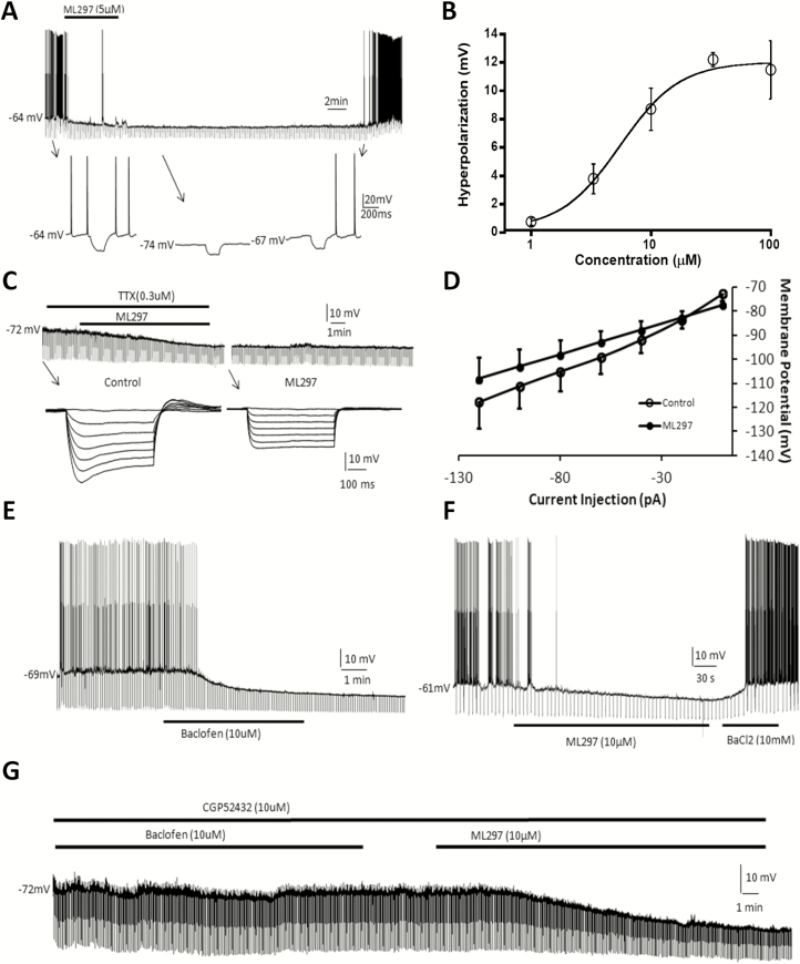
GIRK channel direct activation with ML297 causes hyperpolarization and a decrease in input resistance in hypothalamic hypocretin neuron and hippocampal CA1 neurons. (A) ML297 (5 µM) caused a long-lasting hyperpolarization, decreased input resistance, and blocked spontaneous firing of action potentials of a hypocretin neuron in the hypothalamic brain slice. Lower inserts indicated by arrows show time-expanded traces. (B) The concentration-dependent response curve of hyperpolarization by ML297 reveals an EC50 of 5.4 µM (each data point represents n = 3–5 cells). (C) In the presence of tetrodotoxin (0.3 µM), ML297 (5 µM) induced postsynaptic hyperpolarization and a decrease in input resistance. Lower inserts indicated by arrows show time-expanded traces. (D) Membrane potential changes in response to a series of current injections revealed a current-voltage relationship where the intersection point of the two current–voltage curves in the presence and absence of ML297 indicated a reversal potential of −84 mV (n = 11), which is close to the K+ equilibrium potential and the experimental condition. (E) R-baclofen (10 µM) hyperpolarized a hippocampal pyramidal CA1 neuron by −12 mV, decreased input resistance to 40% of control level, and blocked spontaneous firing of action potentials. (F) ML297 (10 µM) induced similar postsynaptic responses as baclofen in another hippocampal CA1 neuron, which was reversibly blocked by BaCl2 (10 mM), a general K+ channel blocker. (G) In another CA1 neuron, in the presence of the GABAB antagonist CGP52432 and tetrodotoxin (TTX, 0.3 µM), R-baclofen (10 µM) did not induce any postsynaptic responses. Subsequent application of 10 µM ML297 induced responses of hyperpolarization and an increase in input resistance. Hyperpolarizing current pulses (−0.3 nA, 500 ms) were delivered every 5 s throughout the experiment in all recorded neurons which had resting potentials between −50 and −65 mV.

