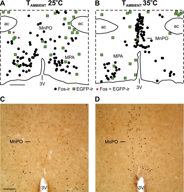
Figure 6.
An acute increase in TAMBIENT activated Fos in the MnPO but not in preoptic NK3R neurons. (A, B) Maps of NK3R (EGFP-ir neurons, green squares), Fos-ir neurons (black circles), and dual labeled neurons (red stars) in a representative OVX mouse exposed to TAMBIENT of (A) 25°C and (B) 35°C. (C, D) Photomicrographs showing the increase in Fos-ir neurons in the MnPO (black; nuclear stain) at a TAMBIENT of 35°C. EGFP-ir neurons not expressing Fos can also be seen (brown; cytoplasmic stain). (A) Scale bar, 250 µm for (A) and (B). (C) Scale bar, 100 µm for (C) and (D). 3V, third ventricle; ac, anterior commissure.

