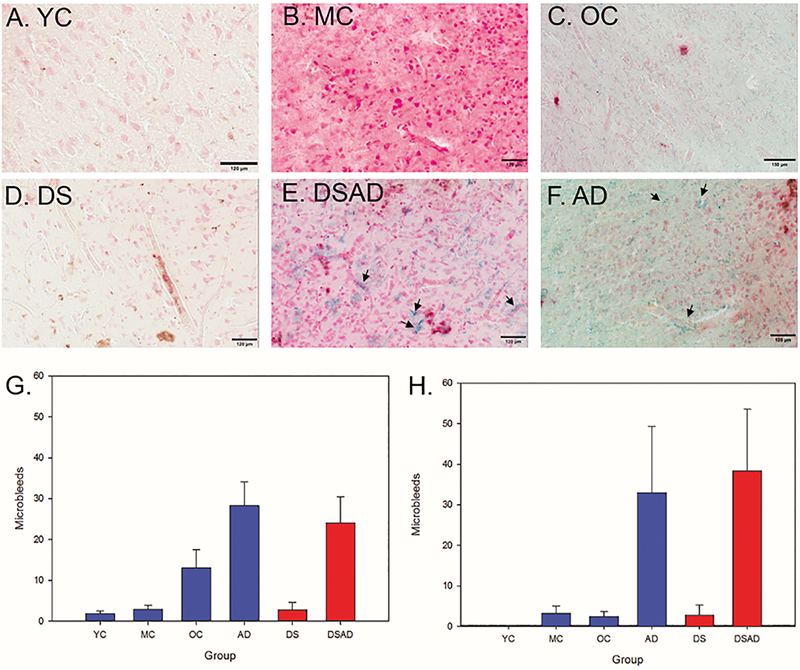
Figure 3: Microbleeds increase with age in the FCTX and OCTX.
Prussian blue staining in our control (A-C) and non-control (D-F) groups, with positive MB labeling marked with arrows; all scale bars represent 120 µm. (A) YC (Age=39), (B) MC (age=56), (C) OC (age=76), (D) DS (age=2), (E) DSAD (age=57), (F) AD (age=76). MB counts were highest in the AD and DSAD group for both the FCTX (G) and OCTX (H). Arrows highlight areas where there are MBs.

