Figure 3.
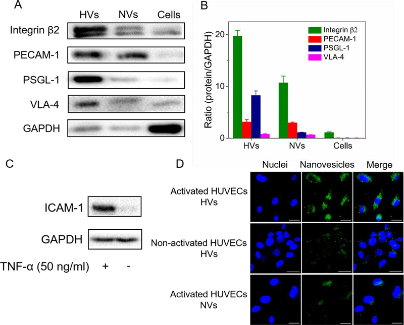
In vitro studies on the binding of HVs to endothelial cells. (A) Western blot of integrin β2, PECAM-1, PSGL-1 and VLA-4 on HL-60 nanovesicles (HVs), non-differentiated HL-60 nanovesicles (NVs) and differentiated HL-60 cells lysis (Cells). (B) Ratios of protein expression levels in HVs, NVs and cells lysis relative to GAPDH (n=3). (C) ICAM-1 expression in normal HUVECs and inflamed HUVECs induced by TNF-α. (D) Fluorescence confocal images show the binding of DiO-labeled nanovesicles (green) to inflamed HUVECs stained by DAPI (blue). (Scale bar=100 μm). HUVECs were activated by TNF-α, followed by incubation with HVs or NVs.

