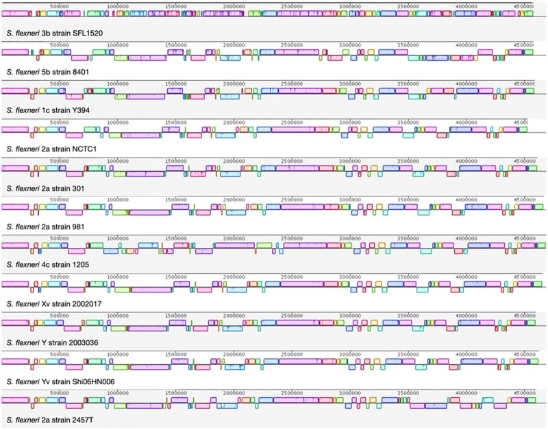
Fig. 2.
—Whole-genome alignment of S. flexneri strains. The horizontal panels represent the genome sequences from top to bottom: S. flexneri 3b strain SFL1520, S. flexneri 5b strain 8401, S. flexneri 1c strain Y394, S. flexneri 2a strain NCTC1, S. flexneri 2a strain 301, S. flexneri 2a strain 981, S. flexneri 4c strain 1205, S. flexneri Xv strain 20021017, S. flexneri Y strain 2003036, S. flexneri Yv strain Shi06HN006, and S. flexneri 2a strain 2457T. Each colored block refers to a shared synteny among the compared strains. Blocks above and below their respective line depict the orientation of the genomic region with respect to SFL1520. The genomes were added sequentially for comparison based on the phylogenetic distances.

