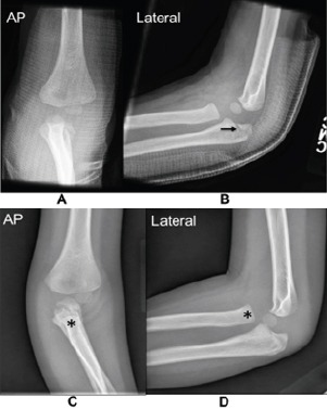
Figure 1.

Anteroposterior (AP) (a) and lateral (b) radiographs of the left elbow 1 day after the injury demonstrate a minimally displaced intra-articular olecranon fracture (arrow) with an overlying fiberglass cast. 5 weeks after the injury, AP (c) and lateral (d) radiographs reveal a healing, non-displaced olecranon fracture with callus formation and bony remodeling in the setting of a dislocation of the radial head (asterisk) medially and anteriorly.
