
Figure 2.
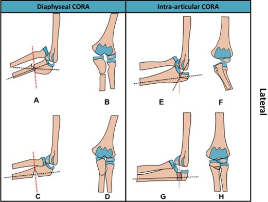
The center of rotational angulation (CORA-dashed red lines) in a lateral (a) and anteroposterior (AP) (b) view of the most common Bado type I (anterior) Monteggia fracture-dislocation displayed with a lateral (c) and AP (d) views of the ulnar osteotomy at the diaphyseal CORA. By contrast, lateral (e) and AP (f) views of the intra-articular Monteggia variant suggest the need for an intra-articular osteotomy to address the CORA and reduce the radial head as shown in the post-operative lateral (g) and AP (h) views. Iliac crest allograft (darker brown shade) was used in the presented intra-articular case. Anatomical axis (dashed black lines) of the proximal and distal ulnar fragment is shown to intersect at the CORA.