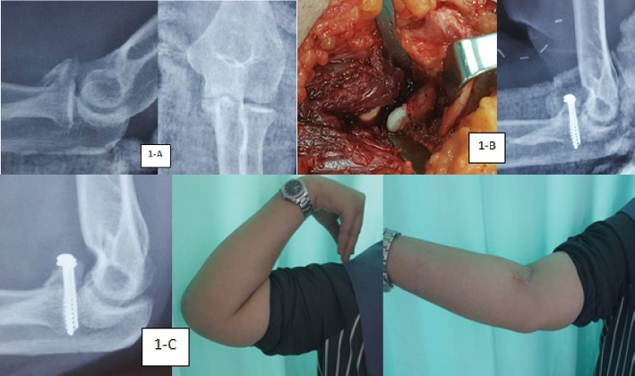
Figure 1.

(a) The pre-operative plain radiographs (lateral and anteroposterior views) of the right elbow. (b) The intraoperative identification of the coronoid process and post-operative plain radiograph of the elbow after the internal fixation with two cancellous lag screws and washer. (c) The full range of movement of the patient’s right elbow and bony union of the coronoid process 3-month post-operation.
