
Figure 4. MRP1 Expression Collaterally Sensitizes to Ferroptosis.
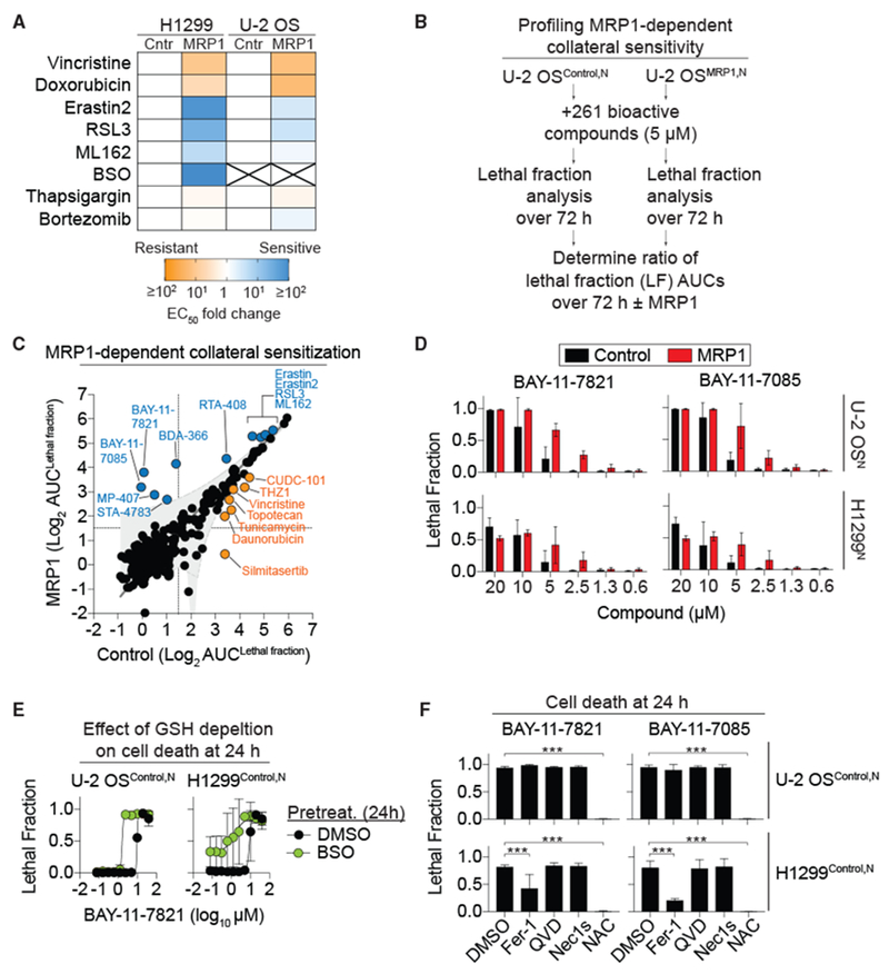
(A) Summary of lethal compound EC50 fold changes for control (Cntr) and MRP1-overexpressing (MRP1) cells. X, EC50 values could not be computed. See also Table 1.
(B) Outline of the collateral sensitivity profiling experiment in Control and MRP1-overexpressing cells in response to 261 bioactive compounds using STACK.
(C) Results of the comparative analysis of cell death ± MRP1. Each dot represents a single compound. MRP1 overexpression had no effect (black), reduced sensitivity (yellow), or increased sensitivity (blue) to compound-induced cell death.
(D) Cell death at 48 h in Control and MRP1-overexpressing cells determined using STACK.
(E) BAY-11-7821-induced cell death determined using STACK ± buthionine sulfoximine (BSO) pretreatment (200 μM, 24 h).
(F) Cell death determined using STACK. Conditions were: BAY-11-7821 or BAY-11-7085 (both 20 μM) ± Fer-1 (2 μM), Q-VD-OPh (25 μM), Nec-1 s (1 μM), or N-acetylcysteine (NAC, 1 mM). Data were analyzed by one-way ANOVA with ***p < 0.001.
Data in (D)–(F) represent means ± SDs for three independent experiments.