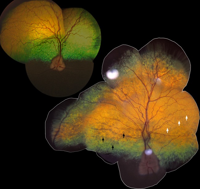
Fig 1. Retinal morphology in vivo in canine Stargardt disease.
The tapetal fundus of the right eye from an 11-year-old unaffected Labrador retriever (upper left; LAB27) and a 10-year-old affected dog (lower right; LAB4). Black arrows show areas with abnormal, grayish, hyporeflective appearance and white arrows indicate attenuation of the retinal blood vessels.

