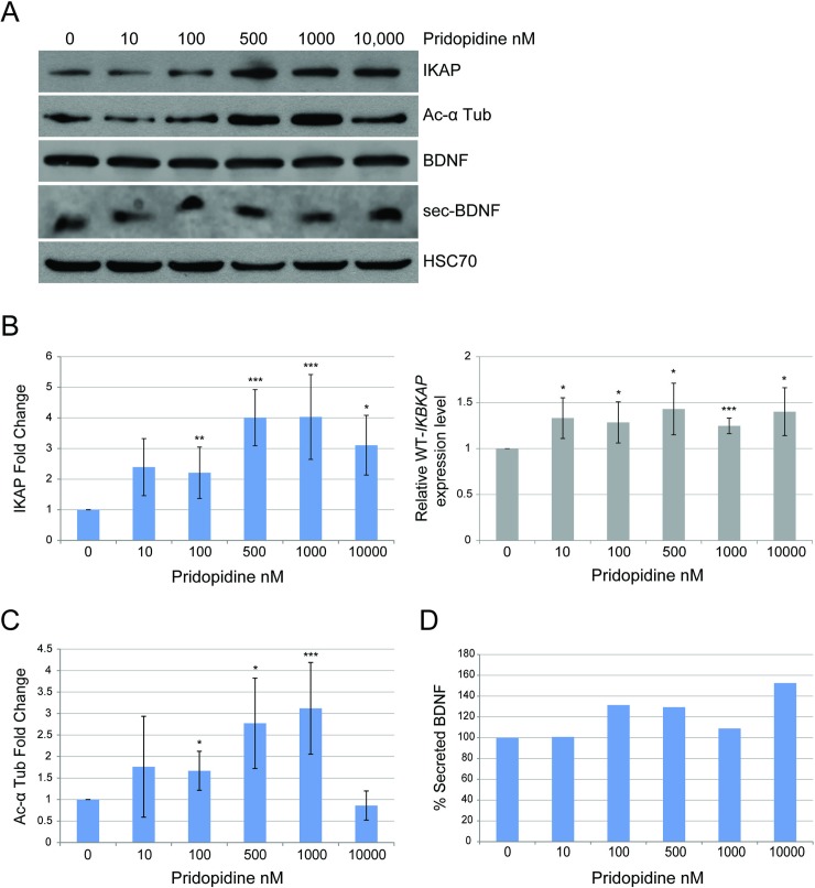
Fig 2. Pridopidine elevates IKAP protein level and affects acetylated α-tubulin levels and BDNF secretion in FD cells.
(A-D) FD cells were treated with 0, 10, 100, 500, 1000, and 10,000 nM of pridopidine. Control treatment was done with vehicle only. (A) Western blotting of FD cell lysates with and without pridopidine treatment for 10 days. Blot was probed using anti-IKAP, anti-acetylated α-tubulin, anti-BDNF, and anti-HSC-70 antibodies. HSC-70 was analyzed as a protein-loading control. (B) Left panel: Fold change levels of IKAP relative to control analyzed after a 10-day treatment; levels were normalized to HSC70. Right panel: IKBKAP expression levels relative to control analyzed by qRT-PCR from RNA extracted from FD cells after 5 days of treatment. (C) Fold change levels of acetylated α-tubulin relative to control normalized to HSC70 analyzed after 10 days of treatment. (D) Quantification of percent BDNF secretion after a 72-hour incubation with pridopidine. All quantifications were done using FusionCapt software. Asterisks denote statistically significant differences (*P ≤ 0.05, **P ≤ 0.01, and ***P ≤ 0.005) relative to control (vehicle only); Student’s t-test.

