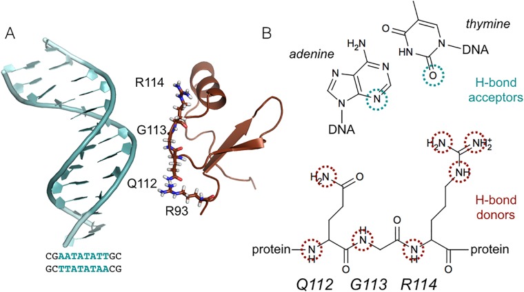
Fig 1. System details.
(A) Snapshot of the initial unbound configuration. The protein (brown) and DNA (light blue) are shown in cartoon representation. The QGR motif (residues 112-114) and R93 are shown as sticks. (B) Scheme indicating the hydrogen bond partners involved in the binding of H-NS to DNA. The donors are provided by the QGR motif in H-NS, highlighted by brown circles. The acceptors are located on the bases at the minor groove side of the DNA, indicated by blue circles.

