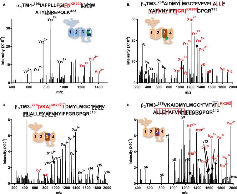
Fig 3. Fragmentation spectra of KK200- and KK202-photolabeled GABAA receptor peptides.
(A) An α1-TM4 peptide (m/z = 898.002, z = 4) is photolabeled by KK200 at N408; (B) A β3-TM3 peptide (m/z = 1,188.352, z = 4) is photolabeled by KK200 at [308GR309]. (C and D) β3-TM3 peptides (m/z = 811.453, z = 6) are photolabeled by KK202 at 226VKA228 (panel C) and L294 (panel D). Fragment ions labeled in red contain the neurosteroid adduct. The C* indicates that the cysteine is alkylated by NEM or NEM + DTT. The photolabeled residues shown in panels A–D were all observed in three replicate experiments. The inset schematic of a GABAA receptor subunit in each panel indicates the approximate location of the residues labeled by KK200 (green) and KK202 (blue). The numerical data are included in S3 Data. DTT, 1, 4-dithiothreitol; GABAA, gamma amino-butyric acid Type A; NEM, N-ethylmaleimide; TM, transmebrane helix.

