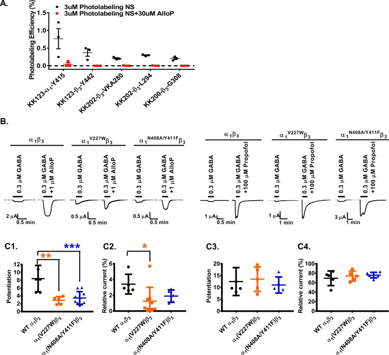
Fig 4. The α1 subunit is critical for neurosteroid modulation of α1β3 GABAA receptors.
(A) Photolabeling with 3 μM KK123, KK200, and KK202 (black) is prevented by labeling in the presence of 30 μM allopregnanolone (red). Each circle represents a replicate experiment, and the bars represent mean ± SD, n = 3. (B) Sample current traces showing modulation of WT α1β3, α1V227Wβ3 and α1N408A/Y411Fβ3 GABAA receptors by allopregnanolone or propofol. (C1) Allopregnanolone potentiation of GABA-elicited currents is reduced in α1V227Wβ3 (orange) and α1N408A/Y411Fβ3 (blue) receptors compared to WT (black). Potentiation is given as the ratio of peak responses to GABA + steroid to GABA alone. Potentiation value of 1 indicates no potentiation by neurosteroids. (C2) Direct activation of α1β3 GABAA receptors by allopregnanolone is reduced in α1V227Wβ3 receptors (orange). Direct activation is given in percentage of the peak response to steroid to peak response to saturating GABA + 100 μM propofol. (C3) Potentiation of GABA-elicited currents by propofol, or (C4) direct activation of the receptor in the presence of propofol is not affected by the α1V227W or α1N408A/Y411F mutations. Each data point represents a replicate experiment. Bars show the mean ± SD (n = 6–8). Data were compared by a one-way analysis of variance followed by a Dunnett’s multiple comparison tests of the means. ***p < 0.001; **p < 0.01; *p < 0.05. The numerical data are included in S4 Data. AlloP, allopregnanolone; GABAA, gamma amino-butyric acid Type A; WT, wild-type.

