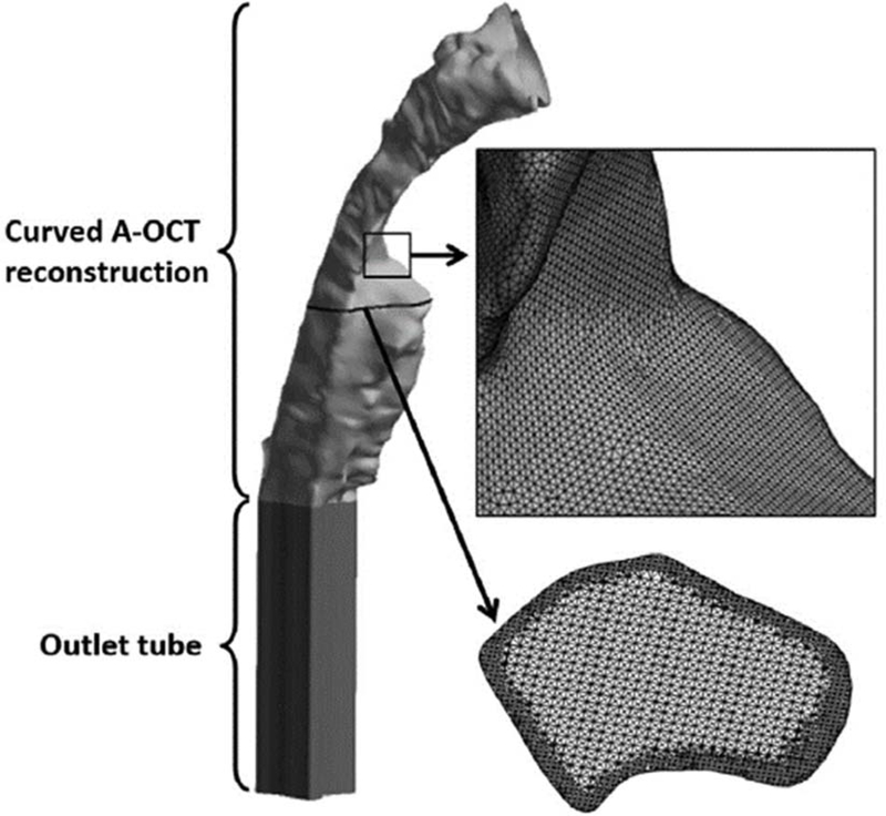
Fig. 3.
LR-OCT airway reconstruction for one neck curvature in one subject with an outlet tube added for numerical stability and illustrations of the computation mesh. Small black box shows an area of enlargement (indicated by short arrow) illustrating the density of the surface mesh. Horizontal black line shows the level of an enlarged axial cross section (indicated by long arrow) illustrating the interior mesh.

