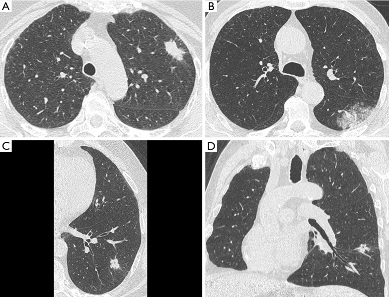
Figure 2.
Morphological heterogeneity of STAS positive lung cancer as displayed by pre-surgical HRCT. (A) Solid nodule with peripheral location and minor subtle spiculations (pT3N0); (B) subsolid mass in the apical segment of left lower lobe (pT1bN0); (C) solid nodule with central hyperlucency on HRCT, defined as cavitation or pseudocavitation (axial reformatting); (D) solid nodule with central hyperlucency on HRCT, defined as cavitation or pseudocavitation (oblique coronal reformatting). STAS, spread through air spaces.

