Figure 1.

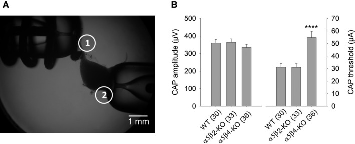
CAP amplitudes do not differ between WT, α5β2‐KO, and α5β4‐KO SCG ganglia. (A) Image showing the experimental setup for recording CAPs in the isolated mouse SCG. The suction electrode for stimulating the preganglionic sympathetic nerve is indicated as electrode 1, and the suction electrode for recording the postganglionic internal carotid nerve is indicated as electrode 2. (B) Summary of CAP amplitude (left) and the stimulus threshold for inducing a CAP (right) on WT, α5β2‐KO, and α5β4‐KO ganglia. CAP amplitude induced by supramaximal stimulation at 0.033 Hz was similar between genotypes (one‐way ANOVA, F 2,96 = 0.75; P = 0.48). In contrast, the stimulus threshold for eliciting a discernible CAP with an amplitude of 15–20 μV was significantly higher in α5β4‐KO compared to both WT and α5β2‐KO mice (one‐way ANOVA, followed by Bonferroni's post hoc test, P < 0.0001). In this and subsequent figures, summary data are presented as the mean ± SEM. See Figure 4 for example CAP recordings.
