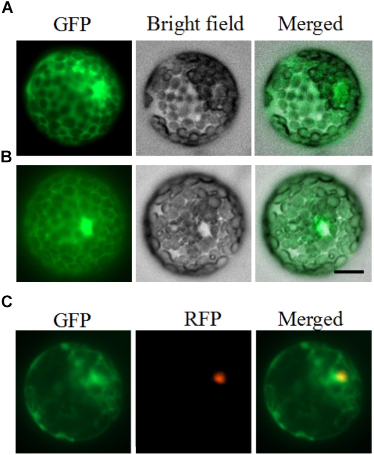
FIGURE 2.

Subcellular localization of PhDHS. Petunia protoplasts were transfected with a construct carrying GFP or PhDHS-GFP under the control of CaMV 35S promoter to assess subcellular localization. (A) Expression of PhDHS-GFP fusion protein. (B) Expression of GFP protein. (C) Subcellular localization of PhDHS-GFP with EOBI-RFP as makers. All images were captured with a confocal laser scanning system. Scale bar: 5 μm.
