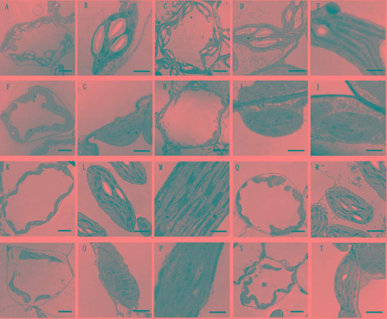
FIGURE 5.
Effects of PhDHS silencing on chloroplast development. (A–J) Chloroplast ultrastructure of mesophyll chloroplasts in the S1 (A,B,F,G) and S2 (C–E,H–J) stages of leaves in 5-week-old pTRV2 empty vector-infected (A–E) and PhDHS-silenced (F–J) plants. (A,C,F,H) Overviews depicting the chloroplast morphology and arrangement within the cells (scale bar, 5 μm). (B,D,G,I) Inside chloroplast views depicting thylakoid arrangement (scale bar, 1 μm). (E,I) Higher magnification depicting the grana stacking (scale bar, 0.5 μm). (K–P) Chloroplast ultrastructure of mesophyll chloroplasts in the S3 stage of leaves in 5-week-old pTRV2 empty vector-infected (K–M) and PhDHS-silenced (N–P) plants. (K,N) Overviews depicting the chloroplast morphology and arrangement within the cells (scale bar, 5 μm). (L,O) Inside chloroplast views depicting thylakoid arrangement (scale bar, 1 μm). (M,P) Higher magnification depicting the grana stacking (scale bar, 0.5 μm). (Q–T) Chloroplast ultrastructure in the leaves of S4 stage in pTRV2 empty vector-infected (Q,R) and PhDHS-silenced (S,T) plants. (Q,S) Overviews depicting the chloroplast morphology and arrangement within the cells (scale bar, 5 μm). (R,T) Inside chloroplast views depicting thylakoid arrangement (scale bar, 1 μm).

