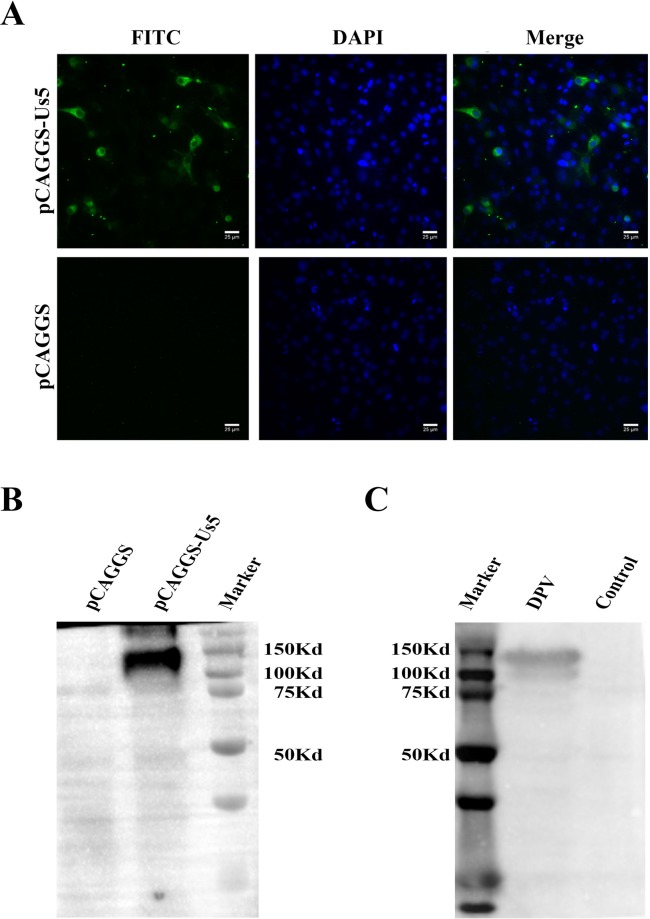
Figure 7.
Expression of the Us5 protein in DEF cells. (A) Indirect immunofluorescence of DPV Us5 in DEF cells. Costaining was performed with a rabbit anti-DPV-Us5 polyclonal antibody and a fluorescein-conjugated goat anti-rabbit IgG antibody. (B) Western blotting of DPV Us5 in DEF cells. A rabbit anti-DPV-Us5 polyclonal antibody was used as the primary antibody. A goat anti-rabbit IgG (H + L)-HRP antibody was used as the secondary antibody. Noninfected cells were used as controls. (C) Western blotting of pCAGGS-Us5 in DEF cells. A duck anti-DPV polyclonal antibody was used as the primary antibody. A goat anti-duck IgG (H + L)-HRP antibody was used as the secondary antibody. Cells transfected with the pCAGGS plasmid were used as the controls.

