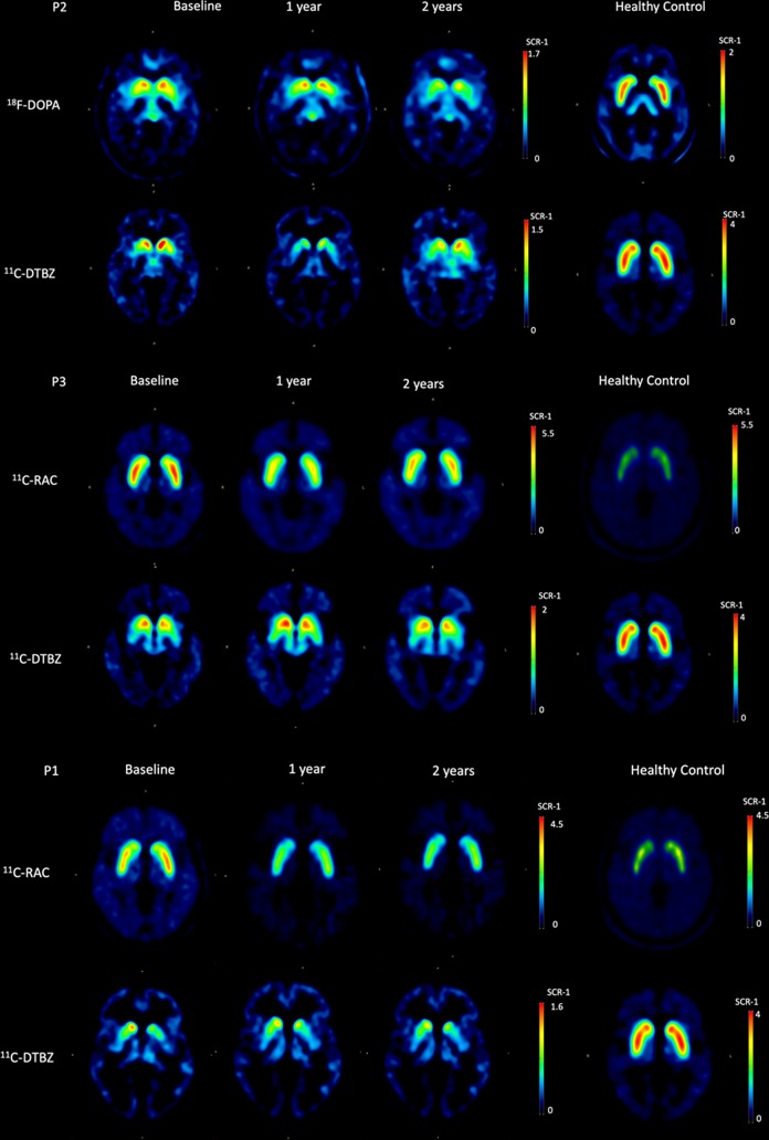
Figure 3.
PET molecular imaging. Representative (P2 and P3) axial RAC, FDOPA and DTBZ PET images of healthy controls and PD patients in their OFF condition for in vivo pre- and post-synaptic assessment of the state of their nigrostriatal dopaminergic system (putamina and caudate nuclei) at baseline, and 1- and 2 years after surgery. P1 is included as an outlier notable for having a decrease in RAC uptake with lack of improvement in motor performance.

