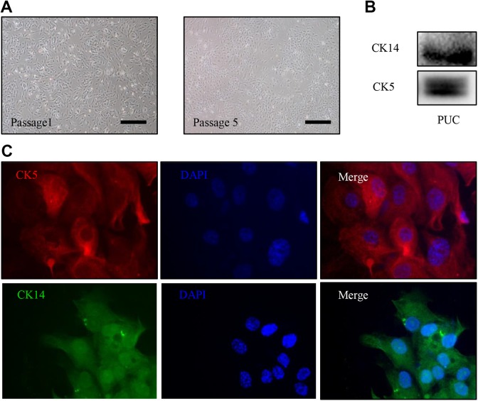
Fig 1.
Expanded PUC cells express urothelial stem/progenitor cell markers. (A) The representative images of PUC cells at passage 1 and 5 are shown. (B) Western blot analysis for CK5 and CK14 expression was performed on passage 3 PUC cells. (C) Immunofluorescence staining of passage 3 PUC cells was carried out with antibodies against CK5 (red) and CK14 (green), nuclei stained with DAPI, 400× magnification. Scale bars represent 50 µm.
DAPI: 4′6-diamidino-2-phenylindole; PUC: porcine urothelial cell.

