FIG 5.
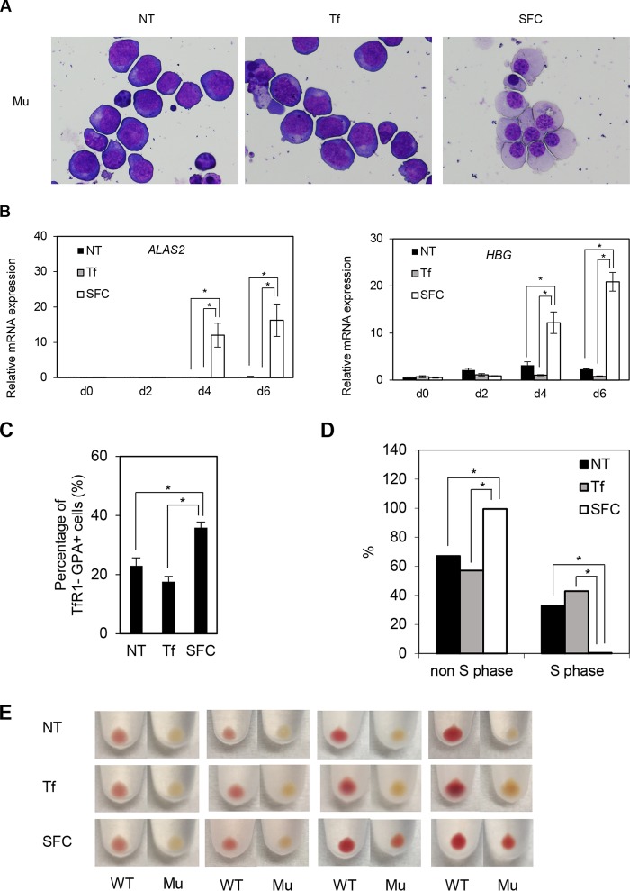
SFC promotes erythroid differentiation of XLSA clone cells. (A) May-Giemsa staining of XLSA clone cells cocultured with OP9 cells for 6 days. NT, nontreated; Tf, iron-saturated transferrin; SFC, sodium ferrous citrate. (B) Quantitative RT-PCR analysis for ALAS2 and HBG expression in XLSA clone cells that were cocultured with OP9 cells for 2, 4, and 6 days. Values presented are relative to those for GAPDH mRNA. Data represent averages from three independent experiments and are expressed as means ± standard deviations. *, P < 0.05. (C) FACS analysis of XLSA clone cells cocultured with OP9 cells for 6 days. The percentages of TfR1-negative, GPA-positive fractions are summarized. Data represent averages from three independent experiments and are expressed as means ± standard deviations. *, P < 0.05. (D) BrdU incorporation assay by FACS. BrdU-positive and -negative fractions denote S phase and non-S phase, respectively. Data represent averages from three independent experiments and are expressed as means ± standard deviations. *, P < 0.05. (E) Cell pellets of cocultured HiDEP cells. Cell pellets of wild-type HiDEP cells and XLSA clone cells cocultured with OP9 cells for 2, 4, and 6 days. Representative data from at least two independent experiments are shown.

