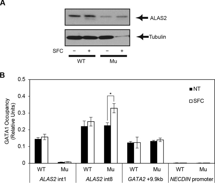
FIG 6.

Increased ALAS2 expression in the ring sideroblasts. (A) Western blot analysis to detect endogenous ALAS2 protein from wild-type HiDEP cells and XLSA clone cells by coculturing with OP9 cells in the presence and absence of SFC. Tubulin was used as a loading control. (B) Quantitative ChIP analysis to detect endogenous GATA-1 chromatin occupancy in wild-type HiDEP cells and in XLSA clone cells cocultured with OP9 cells for 6 days in the presence or absence of SFC. GATA-2 + 9.9 kb, which is the GATA switch site during erythroid differentiation, and the NECDIN promoter were used as positive and negative controls, respectively (6). Data are expressed as means ± standard deviations (n = 3). *, P < 0.05. Average negative-control (IgG) signal did not exceed 0.003.
