FIG 8.
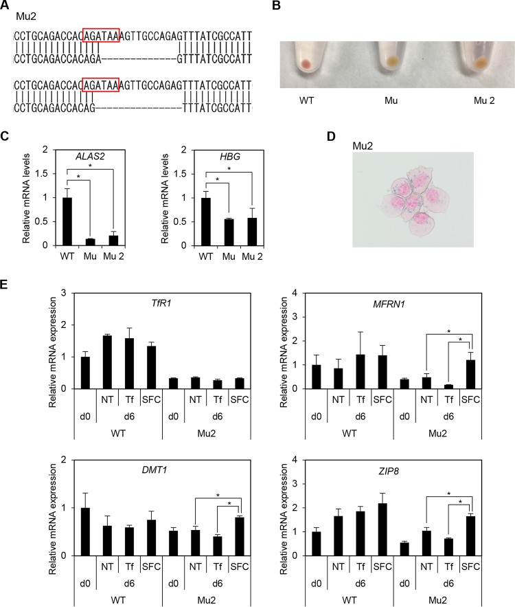
Another clone of ALAS2-mutated HiDEP cells (Mu2). (A) Sequence analysis of another XLSA clone (Mu2). Each allele showed 13- and 15-bp deletions involving GATA binding motifs. (B) Cell pellets of mutant clones. Mu is presented in Fig. 3. (C) Quantitative RT-PCR analysis for ALAS2 and HBG expression in wild-type HiDEP cells and in XLSA clone cells. Values presented are relative to those of GAPDH mRNA. Data represent averages from three independent experiments and are expressed as means ± standard deviations. *, P < 0.05. (D) Prussian blue staining of Mu2 cocultured with OP9 cells supplemented with SFC. (E) Quantitative RT-PCR analysis for TfR1, MFRN1, DMT1, and ZIP8 in wild-type HiDEP cells and in another XLSA clone (Mu2) that were cocultured with OP9 cells for 0 and 6 days. NT, nontreated; Tf, iron-saturated transferrin; SFC, sodium ferrous citrate. Values presented are relative to those of GAPDH mRNA. Data represent averages from three independent experiments and are expressed as means ± SD. *, P < 0.05.

